Skip to Content
MilliporeSigma
All Photos(4)

Key Documents

SAB4200767

Sigma-Aldrich

Anti-Cathepsin D antibody, Mouse monoclonal

clone CTD-19, purified from hybridoma cell culture

Synonym(s):

Anti-CTSD, cleaved into the following 2 chains: Cathepsin D light chain and Cathepsin D heavy chain

Sign Into View Organizational & Contract Pricing


About This Item

UNSPSC Code:
12352203
NACRES:
NA.41

biological source

mouse

Quality Level

antibody form

purified from hybridoma cell culture

antibody product type

primary antibodies

clone

CTD-19, monoclonal

form

buffered aqueous solution

species reactivity

human, rabbit

concentration

~1.0 mg/mL

technique(s)

immunoblotting: 2-4 μg/mL using human breast cancer MCF7 cell line
immunofluorescence: 5-10 μg/mL using HeLa cells
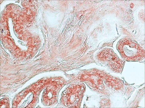
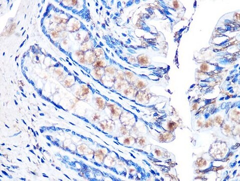
immunohistochemistry: 10-20 μg/mL using heat-retrieved formalin-fixed, paraffin-embedded human liver sections

isotype

IgG2a

UniProt accession no.

shipped in

dry ice

storage temp.

−20°C

target post-translational modification

unmodified

Gene Information

human ... CTSD(1509)

General description

Anti-Cathepsin D antibody, Mouse monoclonal (mouse IgG2a isotype) is derived from the CTD-19 hybridoma, produced by the fusion of mouse myeloma cells and splenocytes from BALB/c mouse immunized with cathepsin D purified from human liver. Cathepsin D (CatD or CTSD) is synthesized in the rough endoplasmic reticulum as pre-proprotein. After removal of signal peptide, the pro-CatD is targeted to endosomes to form an active, ~48 kDa, single-chain intermediate then to the lysosomes to form the fully active mature protease, composed of a ~30 kDa heavy chain and a ~14 kDa light chain.

Immunogen

Cathepsin D Purified from human liver

Application

Anti-Cathepsin D antibody, Mouse monoclonal has been used in:
  • immunoblotting
  • immunohistochemistry
  • immunofluorescence

Biochem/physiol Actions

CatD plays numerous physiological functions in the cells including metabolic degradation of intracellular proteins and the activation of enzymatic precursors. In the central nervous system, CatD is particularly important for the control of neuronal homeostasis, cell migration and interneuron communication. CatD-mediated proteolysis mediates the degradation of unfolded/oxidized protein aggregates in lysosome. The level of CatD synthesized by the cells is increased in response to mitogenic signals from estrogen, epidermal growth factor (EGF), fibroblast growth factor (FGF), and insulin like growth factor-I (IGF- I). The ability of tumor cells to invade the extracellular matrix has been attributed to cathepsins released by tumor cells or associated with its plasma membrane.

Physical form

Solution in 0.01 M phosphate buffered saline, pH 7.4, containing 15 mM sodium azide.

Not finding the right product?  

Try our Product Selector Tool.

Storage Class Code

10 - Combustible liquids

Flash Point(F)

Not applicable

Flash Point(C)

Not applicable


Certificates of Analysis (COA)

Search for Certificates of Analysis (COA) by entering the products Lot/Batch Number. Lot and Batch Numbers can be found on a product’s label following the words ‘Lot’ or ‘Batch’.

Already Own This Product?

Find documentation for the products that you have recently purchased in the Document Library.

Visit the Document Library

M J Duffy et al.
Clinical chemistry, 37(1), 101-104 (1991-01-01)
Cathepsin D (CD, EC 3.4.23.5) is a lysosomal protease induced by estrogen in certain estrogen receptor (ER)-positive breast cancer cell lines but produced constitutively by ER-negative cell lines. Our aims in this investigation were to study the distribution of CD
Nuclear cathepsin D enhances TRPS1 transcriptional repressor function to regulate cell cycle progression and transformation in human breast cancer cells
Bach AS, et al.
Oncotarget, 6(29), 28084-28084 (2015)
Cathepsin D as a therapeutic target in Alzheimer's disease.
Di Domenico F, et al.
Expert Opinion on Therapeutic Targets, 20(12), 1393-1395 (2016)
The role of cathepsin D in the pathogenesis of human neurodegenerative disorders
Vidoni C, et al.
Medicinal Research Reviews, 36(5), 845-870 (2016)
Veronika Stoka et al.
Ageing research reviews, 32, 22-37 (2016-04-30)
Lysosomes and lysosomal hydrolases, including the cathepsins, have been shown to change their properties with aging brain a long time ago, although their function was not really understood. The first biochemical and clinical studies were followed by a major expansion

Our team of scientists has experience in all areas of research including Life Science, Material Science, Chemical Synthesis, Chromatography, Analytical and many others.

Contact Technical Service