Skip to Content
MilliporeSigma
All Photos(2)

Key Documents

S2657

Sigma-Aldrich

Monoclonal Anti-S-100 (β-Subunit) antibody produced in mouse

clone SH-B4, ascites fluid

Synonym(s):

Anti-NEF, Anti-S100, Anti-S100-B, Anti-S100beta

Sign Into View Organizational & Contract Pricing


About This Item

MDL number:
UNSPSC Code:
12352203
NACRES:
NA.41

biological source

mouse

Quality Level

conjugate

unconjugated

antibody form

ascites fluid

antibody product type

primary antibodies

clone

SH-B4, monoclonal

contains

15 mM sodium azide

species reactivity

sheep, bovine, goat, pig, feline, rabbit, rat, canine, human

technique(s)

immunohistochemistry (formalin-fixed, paraffin-embedded sections): suitable (: suitable)
indirect ELISA: suitable (:suitable)
western blot: 1:100-1:200 ( (using S-100B Protein, Bovine Brain))

isotype

IgG1

UniProt accession no.

shipped in

dry ice

storage temp.

−20°C

target post-translational modification

unmodified

Gene Information

human ... S100B(6285)
rat ... S100b(25742)

General description

Monoclonal Anti-S-100 (b-Subunit) (mouse IgG1 isotype) is derived from the SH-B4 hybridoma produced by the fusion of mouse myeloma cells and splenocytes from immunized BALB/c mice. S-100 is a set of small, highly acidic, thermolabile dimer proteins of ~20 kDa, widely distributed in different tissues.
S-100 (β-Subunit) is a calcium-binding protein, that belongs to S100 family of calcium-binding proteins. It is expressed in melanocytes. It is located on human chromosome 21q22.3.

Specificity

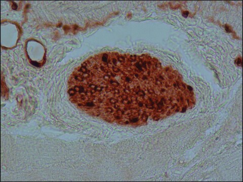
Recognizes an epitope located on the β chain (i.e. in S-100a and S-100b) but not on the α chain of S-100 (i.e. in S-100a and S-100ao). In ELISA, recognition of S-100 subunit by clone SH-B4 is Ca2+-dependent. The product does not react with other members of the EF-hand family such as, calmodulin, parvalbumin, intestinal calcium-binding protein and myosin light chain. In immunohistochemistry, the antibody detects normal and neoplastic S-100 β subunit-containing cells (e.g. Schwann cells, chondrocytes, melanocytes, and melanotic tumors) in protease-digested, formalin-fixed, paraffin-embedded tissues.

Immunogen

bovine brain S-100b

Application

Applications in which this antibody has been used successfully, and the associated peer-reviewed papers, are given below.
Immunocytochemistry (1 paper)
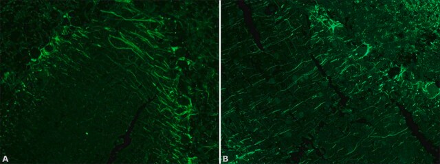
Monoclonal Anti-S-100 (β-Subunit) antibody produced in mouse has been used in immunocytochemistry and immunohistochemistry. It has also been used as a component in serum medium for 5-ethynyl-29-deoxyuridine (EdU), incorporation assay.

Biochem/physiol Actions

S-100 (β-Subunit) acts as a marker of melanocyte cytotoxicity. Reduced expression of S100B is observed in chronic liver diseases. It also acts as a serum marker in endocrine resistant breast cancer.
S-100 is considered to be a cell-growth regulator, but other functions have been suggested, e.g., increasing the membrane permeability to cations under physiologic conditions, stimulation of nucleolar RNA polymerase activity, interaction with the tumor suppressor protein p53 and as a carrier of proteins and free fatty acids in adipocytes.

Disclaimer

Unless otherwise stated in our catalog or other company documentation accompanying the product(s), our products are intended for research use only and are not to be used for any other purpose, which includes but is not limited to, unauthorized commercial uses, in vitro diagnostic uses, ex vivo or in vivo therapeutic uses or any type of consumption or application to humans or animals.

Not finding the right product?  

Try our Product Selector Tool.

Storage Class Code

10 - Combustible liquids

WGK

WGK 3

Flash Point(F)

Not applicable

Flash Point(C)

Not applicable


Choose from one of the most recent versions:

Certificates of Analysis (COA)

Lot/Batch Number

Don't see the Right Version?

If you require a particular version, you can look up a specific certificate by the Lot or Batch number.

Already Own This Product?

Find documentation for the products that you have recently purchased in the Document Library.

Visit the Document Library

Dongsheng Wu et al.
The Journal of neuroscience : the official journal of the Society for Neuroscience, 38(6), 1351-1365 (2017-12-28)
Injury to the peripheral axons of sensory neurons strongly enhances the regeneration of their central axons in the spinal cord. It remains unclear on what molecules that initiate such conditioning effect. Because ATP is released extracellularly by nerve and other
S100 Proteins As an Important Regulator of Macrophage Inflammation
Xia C, et al.
Frontiers in Immunology (2017)
Marked innervation but also signs of nerve degeneration in between the Achilles and plantaris tendons and presence of innervation within the plantaris tendon in midportion Achilles tendinopathy
Spang C, et al.
Journal of musculoskeletal & neuronal interactions, 15(2), 197-197 (2015)
Thermally drawn fibers as nerve guidance scaffolds
Koppes RA, et al.
Biomaterials, 81, 27-35 (2016)
A response to 'Cerebral and extracerebral release of protein S100B in cardiac surgical patients', Snyder-Ramos SA, Gruhlke T, Bauer H, Bauer M, Luntz AP, Motsch J, Martin E, Vahl CF, Missler U, Wiesmann M and Bottiger BW, Anaesthesia 2004; 59: 344-9.
D Janigro
Anaesthesia, 59(11), 1149-1150 (2004-10-14)

Our team of scientists has experience in all areas of research including Life Science, Material Science, Chemical Synthesis, Chromatography, Analytical and many others.

Contact Technical Service