Skip to Content
MilliporeSigma
All Photos(2)

Key Documents

P8232

Sigma-Aldrich

Anti-P2X7 Purinergic Receptor antibody produced in rabbit

affinity isolated antibody, lyophilized powder

Synonym(s):

Anti-P2X₇ Antibody, P2X₇ Receptor Detection, Rabbit Anti-P2X₇

Sign Into View Organizational & Contract Pricing


About This Item

MDL number:
UNSPSC Code:
12352203
NACRES:
NA.41

biological source

rabbit

Quality Level

conjugate

unconjugated

antibody form

affinity isolated antibody

antibody product type

primary antibodies

clone

polyclonal

form

lyophilized powder

species reactivity

human, mouse, rat

technique(s)

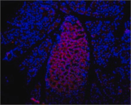
immunohistochemistry: suitable
western blot: 1:200-1:1,000

UniProt accession no.

shipped in

dry ice

storage temp.

−20°C

target post-translational modification

unmodified

Gene Information

human ... P2RX7(5027)
mouse ... P2rx7(18439)
rat ... P2rx7(29665)

General description

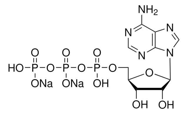
Purinergic receptor P2X7 is an ATP-gated cation channel. It is expressed in retinal pigmented epithelium and neural retina. The gene encoding this protein is localized on human chromosome 12.

Immunogen

Peptide (C)KIRKEFPKTQGQYSGFKYPY, corresponding to amino acid residues 576-595 of rat P2X7 Receptor (Accession Q64663). Intracellular, C-terminus.

Application

Anti-P2X7 Purinergic Receptor antibody produced in rabbit has been used in
  • immunoprecipitation
  • immunofluorescence and confocal microscopy
  • western blotting

Biochem/physiol Actions

Purinergic receptor P2X7 has a role in immunological processes. It has a role during oxidative stress under pathological conditions. The channel is permeable to Na+, Ca2+ and K+. Prolonged activation of the channel under some pathological conditions leads to the passage of large molecules and ultimately cell death. P2X7 may be involved in the release of interleukin-1β (IL-1β) and indirectly initiate an inflammatory cascade.

Physical form

Lyophilized from phosphate buffered saline, pH 7.4, containing 1% bovine serum albumin and 0.05% sodium azide.

Storage and Stability

Prior to reconstitution, store at –20°C. After reconstitution, the stock antibody solution may be stored at 2–8°C for up to two weeks. For extended storage, freeze at -20°C, or below, in working aliquots. Repeated freezing and thawing is not recommended. Storage in "frost-free" freezers is not recommended. Centrifuge all antibody preparations before use (10,000 x g 5 min). Working dilution samples should be discarded if not used within 12 hours.

Other Notes

In order to obtain the best results using various techniques and preparations, we recommend determining the optimal working dilutions by titration.

Disclaimer

Unless otherwise stated in our catalog or other company documentation accompanying the product(s), our products are intended for research use only and are not to be used for any other purpose, which includes but is not limited to, unauthorized commercial uses, in vitro diagnostic uses, ex vivo or in vivo therapeutic uses or any type of consumption or application to humans or animals.

Not finding the right product?  

Try our Product Selector Tool.

Storage Class Code

13 - Non Combustible Solids

WGK

WGK 2

Flash Point(F)

Not applicable

Flash Point(C)

Not applicable


Certificates of Analysis (COA)

Search for Certificates of Analysis (COA) by entering the products Lot/Batch Number. Lot and Batch Numbers can be found on a product’s label following the words ‘Lot’ or ‘Batch’.

Already Own This Product?

Find documentation for the products that you have recently purchased in the Document Library.

Visit the Document Library

Lack of the P2X7 receptor protects against AMD-like defects and microparticle accumulation in a chronic oxidative stress-induced mouse model of AMD.
Carver KA
Biochemical and Biophysical Research Communications, 482(1), 81-86 (2017)
The P2X7 receptor directly interacts with the NLRP3 inflammasome scaffold protein
Alessia Franceschini
Faseb Journal (2015)
Roshni Thakkar et al.
Oxidative medicine and cellular longevity, 2016, 8309031-8309031 (2016-11-16)
17β-Estradiol (E2) is a well-known neuroprotective factor in the brain. Recently, our lab demonstrated that the neuroprotective and cognitive effects of E2 require mediation by the estrogen receptor (ER) coregulator protein and proline-, glutamic acid-, and leucine-rich protein 1 (PELP1).
Autophagy and ATP-induced anti-apoptosis in antigen presenting cells (APC) follows the cytokine storm in patients after major trauma
E. Marion Schneider
Journal of Cell Communication and Signaling, 5, 145-156 (2011)
Qing-Bing Zha et al.
Frontiers in immunology, 7, 597-597 (2016-12-27)
Adenosine triphosphate (ATP) is released by bacteria and host cells during bacterial infection as well as sterile tissue injury, acting as an inducer of inflammasome activation. Previous studies have shown that ATP treatment leads to AMP-activated protein kinase (AMPK) activation.

Our team of scientists has experience in all areas of research including Life Science, Material Science, Chemical Synthesis, Chromatography, Analytical and many others.

Contact Technical Service