Skip to Content
MilliporeSigma
All Photos(3)

Key Documents

P8044

Sigma-Aldrich

Proteinase K from Tritirachium album

≥3.0 unit/mg solid, lyophilized powder

Synonym(s):

Endopeptidase K

Sign Into View Organizational & Contract Pricing


About This Item

CAS Number:
Enzyme Commission number:
EC Number:
MDL number:
UNSPSC Code:
12352204
eCl@ss:
32160410
NACRES:
NA.54

biological source

maize
microbial (T.album
T. ALBUM)

plant seeds (barley)
soybean
yeast

Quality Level

form

lyophilized powder

specific activity

≥3.0 unit/mg solid

mol wt

28.93 kDa

composition

Protein, ≥15% biuret

technique(s)

DNA extraction: suitable

application(s)

diagnostic assay manufacturing

storage temp.

−20°C

Looking for similar products? Visit Product Comparison Guide

Application

Proteinase K is useful for the proteolytic inactivation of nucleases during the isolation of DNA and RNA. It is used for the removal of endotoxins bound to cationic proteins such as lysozyme and ribonuclease A. It is also useful for the isolation of hepatic, yeast, and mung bean mitochondria and is used to determine enzyme localization on membranes. Furthermore, it is used for the treatment of paraffin embedded tissue sections to expose antigen binding sites and for digestion of proteins from brain tissue samples. Product P8044 is provided as a lyophilized powder.
The enzyme from Sigma has been used to degrade complex I (NADH:ubiquinone oxidoreductase) prior to extraction of ubiquinone from Yarrowia lipolytica. It has been used to examine the effect of Proteinase k on Pythium ultimum. It has also been used to maximize the variety of peptide bonds hydrolyzed in the sediment slurry without autolytic production of amino acids from the enzyme itself.
Useful for the proteolytic inactivation of nucleases during the isolation of DNA and RNA.
Removes endotoxins that bind to cationic proteins such as lysozyme and ribonuclease A.
Reported useful for the isolation of hepatic, yeast, and mung bean mitochondria
Determination of enzyme localization on membranes
Treatment of paraffin embedded tissue sections to expose antigen binding sites for antibody labeling.
Digestion of proteins from brain tissue samples for prions in Transmissible Spongiform Encephalopathies (TSE) research.

Biochem/physiol Actions

Proteinase K is highly active towards native proteins. It has a broad specificity and degrades many proteins even in the native state. It mainly cleaves the peptide bond adjacent to the carboxyl group of aliphatic and aromatic amino acids with blocked α-amino groups. The optimum pH is between 7.5-9.0 and the isoelectric point is 8.9. Ca2+ (1-5 mM) is required for activation. Proteinase K is inhibited by diisopropyl fluorophosphate (DFIP), and phenylmethanesulfonyl fluoride (PMSF).
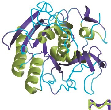
Proteinase K is a stable and highly reactive serine protease. Evidence from crystal and molecular structure studies indicates the enzyme belongs to the subtilisin family with an active-site catalytic triad (Asp39-His69-Ser224). It is stable in a broad range of environments: pH, buffer salts, detergents (SDS), and temperature. In the presence of 0.1-0.5% SDS, proteinase K retains activity and will digest a variety of proteins and nucleases in DNA preparations without compromising the integrity of the isolated DNA.

Unit Definition

One unit will hydrolyze urea-denatured hemoglobin to produce color equivalent to 1.0 μmole of tyrosine per min at pH 7.5 at 37 °C (color by Folin-Ciocalteu reagent).

Pictograms

Health hazardExclamation mark

Signal Word

Danger

Hazard Statements

Hazard Classifications

Eye Irrit. 2 - Resp. Sens. 1 - Skin Irrit. 2 - STOT SE 3

Target Organs

Respiratory system

Storage Class Code

11 - Combustible Solids

WGK

WGK 1

Flash Point(F)

Not applicable

Flash Point(C)

Not applicable

Personal Protective Equipment

dust mask type N95 (US), Eyeshields, Gloves

Certificates of Analysis (COA)

Search for Certificates of Analysis (COA) by entering the products Lot/Batch Number. Lot and Batch Numbers can be found on a product’s label following the words ‘Lot’ or ‘Batch’.

Already Own This Product?

Find documentation for the products that you have recently purchased in the Document Library.

Visit the Document Library

P Bermejo-Alvarez et al.
Biology of reproduction, 79(4), 594-597 (2008-06-27)
It has been reported that the mammalian female could have a preconceptual influence on the sex of her offspring, and it has been hypothesized that this influence could go some way toward accounting for the reported lower fertility following insemination
C Dunne et al.
Microbiology (Reading, England), 146 ( Pt 8), 2069-2078 (2000-08-10)
Stenotrophomonas maltophilia W81 can protect sugar beet against PYTHIUM:-mediated damping-off disease through the production of an extracellular protease. Here, the proteolytic enzyme of W81 was purified by anion-exchange chromatography and characterized as a serine protease. The purified enzyme was fungicidal
Bioavailable amino acids in sediments: A biomimetic, kinetics-based approach
Mayer LM, et al.
Limnology and Oceanography, 40(3), 511-520 (1995)
Stefan Dröse et al.
Biochimica et biophysica acta, 1556(1), 65-72 (2002-09-28)
NADH:ubiquinone oxidoreductase (complex I) is the largest multiprotein complex of the mitochondrial respiratory chain. His-tagged complex I purified from the strictly aerobic yeast Yarrowia lipolytica exhibited electron transfer rates from NADH to n-decylubiquinone of less than 2% when compared to
Enzymes of Molecular Biology
M.M. Burrell
Methods in Molecular Biology, 16, 307-307 (1993)

Articles

Proteinase K (EC 3.4.21.64) activity can be measured spectrophotometrically using hemoglobin as the substrate. Proteinase K hydrolyzes hemoglobin denatured with urea, and liberates Folin-postive amino acids and peptides. One unit will hydrolyze hemoglobin to produce color equivalent to 1.0 μmol of tyrosine per minute at pH 7.5 at 37 °C (color by Folin & Ciocalteu's Phenol Reagent).

Protocols

Proteinase K (EC 3.4.21.64) activity can be measured spectrophotometrically using hemoglobin as the substrate. Proteinase K hydrolyzes hemoglobin denatured with urea, and liberates Folin-postive amino acids and peptides. One unit will hydrolyze hemoglobin to produce color equivalent to 1.0 μmol of tyrosine per minute at pH 7.5 at 37 °C (color by Folin & Ciocalteu's Phenol Reagent).

Our team of scientists has experience in all areas of research including Life Science, Material Science, Chemical Synthesis, Chromatography, Analytical and many others.

Contact Technical Service