Skip to Content
MilliporeSigma
All Photos(6)

Documents

M6319

Sigma-Aldrich

Anti-Mitofusin-2 (N-Terminal) antibody produced in rabbit

affinity isolated antibody, buffered aqueous solution

Synonym(s):

Mitofusin 2 Antibody, Mitofusin 2 Antibody - Anti-Mitofusin-2 (N-Terminal) antibody produced in rabbit, Anti-CMT2A, Anti-CMT2A2, Anti-CPRP1, Anti-KIAA0214, Anti-MARF, Anti-Mfn2

Sign Into View Organizational & Contract Pricing


About This Item

MDL number:
UNSPSC Code:
12352203
NACRES:
NA.41

biological source

rabbit

conjugate

unconjugated

antibody form

affinity isolated antibody

antibody product type

primary antibodies

clone

polyclonal

form

buffered aqueous solution

mol wt

antigen ~86 kDa

species reactivity

mouse, human, rat

technique(s)

immunoprecipitation (IP): 5-10 μg using HeLa human epitheloid carcinoma cell lysate
indirect immunofluorescence: 20-30 μg/mL using differentiated mouse C2 cells
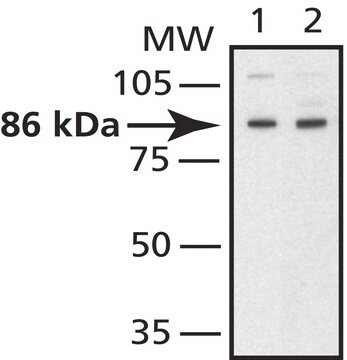
western blot (chemiluminescent): 0.5-1 μg/mL using extracts of rat or mouse brain mitochondria

UniProt accession no.

shipped in

dry ice

storage temp.

−20°C

target post-translational modification

unmodified

Gene Information

human ... MFN2(9927)
mouse ... Mfn2(170731)
rat ... Mfn2(64476)

General description

Mitofusins (Mfn1 and Mfn2) are the mammalian homologs of the Drosophila protein fuzzy onion (Fzo). They are transmembrane GTPases embedded in the outer membrane of mitochondria, essential for fusion of mitochondria in mammalian cells. Mfn1 and Mfn2 form homotypic and heterotypic complexes that are functional for fusion. Mitochondrial fusion is also important for cell growth, mitochondrial membrane potential, respiration, and embryonic development. Mice deficient in either Mfn1 or Mfn2 die in mid-gestation. Mfn2 mutant embryos have a specific and severe disruption of a layer of the placenta. Mitofusin 2 is broadly expressed, with highest expression in heart and skeletal muscle and is induced during myogenesis. Repression of Mfn2 causes morphological and functional fragmentation of the mitochondrial network into independent clusters and reduces mitochondrial membrane potential and glucose oxidation. Thus, Mfn2 is essential for the maintenance of mitochondrial network and controls mitochondrial metabolism. This Mfn2-dependent regulatory mechanism is disturbed in obesity by reduced Mfn2 expression. Mutations in Mitofusin 2 cause Charcot-Marie-Tooth neuropathy type 2A, a neurological disorder that results from degeneration of axons in peripheral nerves.

Specificity

Anti-Mitofusin 2 (N-terminal) antibody recognizes human, rat, and mouse mitofusin 2. Detection of the mitofusin 2 band by immunoblotting is specifically inhibited with the immunizing peptide.
The antibody is specific for N-terminal of mitofusin 2 (~86 kDa)

Immunogen

synthetic peptide corresponding to amino acid residues 38-55 of human mitofusin 2 with C-terminal added cysteine, conjugated to KLH. The corresponding sequence differs by one amino acid in both rat and mouse mitofusin 2.

Application

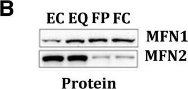
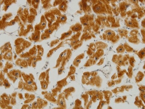
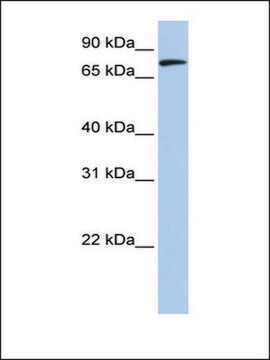
Anti-Mitofusin 2 (N-terminal) antibody is suitable for immunoblotting (~86 kDa), immunoprecipitation, and immunofluorescence applications.
By immunoblotting, a working antibody concentration of 0.5-1 mg/mL is recommended using an extracts of rat and mouse brain mitochondria and a chemiluminescent detection reagent.
By indirect immunofluorescence, a working antibody concentration of 20-30 mg/mL is recommended using differentiated mouse C2 cells.
5-10 mg of the antibody immunoprecipitates mitofusin 2 from HeLa human epithelioid carcinoma cell lysate.
Anti-mitofusion 2 antibody may be used for immunoprecipitation in HeLa cells; immunoblotting in mouse and rat brain mitochondia and immunoflurescence in mouse C2 cells
Applications in which this antibody has been used successfully, and the associated peer-reviewed papers, are given below.
Western Blotting (1 paper)

Physical form

Solution in 0.01 M phosphate buffered saline, pH 7.4, containing 15 mM sodium azide.

Disclaimer

Unless otherwise stated in our catalog or other company documentation accompanying the product(s), our products are intended for research use only and are not to be used for any other purpose, which includes but is not limited to, unauthorized commercial uses, in vitro diagnostic uses, ex vivo or in vivo therapeutic uses or any type of consumption or application to humans or animals.

Not finding the right product?  

Try our Product Selector Tool.

Storage Class Code

12 - Non Combustible Liquids

WGK

WGK 1

Flash Point(F)

Not applicable

Flash Point(C)

Not applicable


Certificates of Analysis (COA)

Search for Certificates of Analysis (COA) by entering the products Lot/Batch Number. Lot and Batch Numbers can be found on a product’s label following the words ‘Lot’ or ‘Batch’.

Already Own This Product?

Find documentation for the products that you have recently purchased in the Document Library.

Visit the Document Library

Cecília García-Pérez et al.
American journal of physiology. Heart and circulatory physiology, 301(5), H1907-H1915 (2011-08-23)
Propagation of ryanodine receptor (RyR2)-derived Ca(2+) signals to the mitochondrial matrix supports oxidative ATP production or facilitates mitochondrial apoptosis in cardiac muscle. Ca(2+) transfer likely occurs locally at focal associations of the sarcoplasmic reticulum (SR) and mitochondria, which are secured
Jonathan P Little et al.
Journal of applied physiology (Bethesda, Md. : 1985), 111(6), 1554-1560 (2011-08-27)
Low-volume high-intensity interval training (HIT) is emerging as a time-efficient exercise strategy for improving health and fitness. This form of exercise has not been tested in type 2 diabetes and thus we examined the effects of low-volume HIT on glucose
Short mitochondrial ARF triggers Parkin/PINK1-dependent mitophagy
Grenier K, et al.
The Journal of Biological Chemistry (2014)
Nicholas R Wawrzyniak et al.
Oncotarget, 7(33), 52695-52709 (2016-07-23)
Fatigue is a symptom of many diseases, but it can also manifest as a unique medical condition, such as idiopathic chronic fatigue (ICF). While the prevalence of ICF increases with age, mitochondrial content and function decline with age, which may
Karl Grenier et al.
The Journal of biological chemistry, 289(43), 29519-29530 (2014-09-14)
Parkinson disease (PD) is a complex neurodegenerative disease characterized by the loss of dopaminergic neurons in the substantia nigra. Multiple genes have been associated with PD, including Parkin and PINK1. Recent studies have established that the Parkin and PINK1 proteins

Our team of scientists has experience in all areas of research including Life Science, Material Science, Chemical Synthesis, Chromatography, Analytical and many others.

Contact Technical Service