Skip to Content
MilliporeSigma
All Photos(2)

Key Documents

L1418

Sigma-Aldrich

Anti-LAMP1 antibody produced in rabbit

affinity isolated antibody, buffered aqueous solution

Synonym(s):

LAMP1 Antibody - Anti-LAMP1 antibody produced in rabbit, Lamp1 Antibody, Anti-CD107a, Anti-LAMPA, Anti-LGP120, Anti-Lysosomal-associated membrane protein 1

Sign Into View Organizational & Contract Pricing


About This Item

MDL number:
UNSPSC Code:
12352203
NACRES:
NA.41

biological source

rabbit

Quality Level

conjugate

unconjugated

antibody form

affinity isolated antibody

antibody product type

primary antibodies

clone

polyclonal

form

buffered aqueous solution

mol wt

antigen ~120 kDa

species reactivity

rat, mouse, human

packaging

antibody small pack of 25 μL

technique(s)

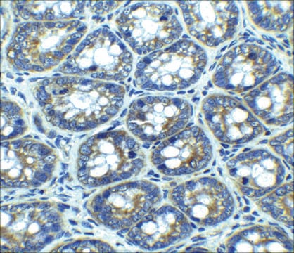
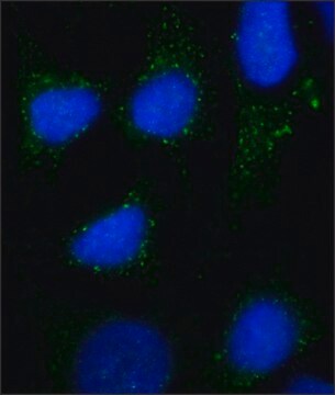
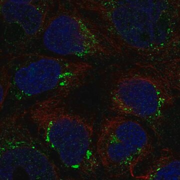
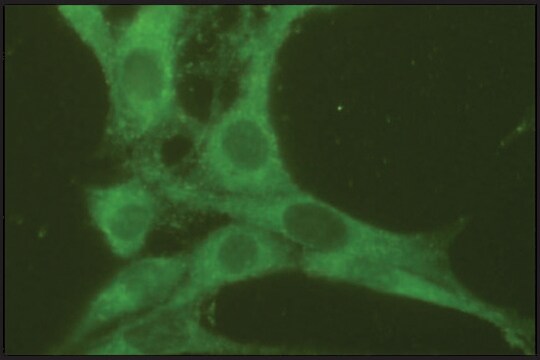
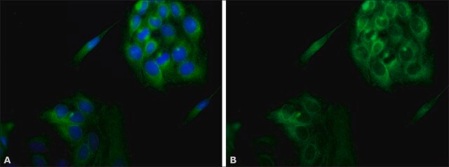
indirect immunofluorescence: 5-10 μg/mL using human HeLa, rat NRK, and mouse NIH3T3 cells

UniProt accession no.

shipped in

dry ice

storage temp.

−20°C

target post-translational modification

unmodified

Gene Information

human ... LAMP1(3916)
mouse ... Lamp1(16783)
rat ... Lamp1(25328)

General description

Lysosome-associated membrane protein 1 (LAMP1), also termed Igp120, is a heavily glycosylated lysosomal membrane protein of about 120 kDa. It consists of a ~40 kDa core polypeptide with O-linked and 18 asparagine-linked oligosaccharide side chains. LAMP1 protein contains a leader sequence, a large intralumenal region consisting of 2 homologous domains separated by a hinge region rich in proline and serine, a 24-amino acid transmembrane region, and a short cytoplasmic tail containing the lysosomal membrane targeting signal. LAMP1 is ubiquitously expressed and highly conserved. It localizes mainly to lysosomes although a small portion is detected on the cell surface. It was found that highly metastatic tumor cells express more LAMP molecules on the cell surface than poorly metastatic cells.
The gene LAMP1 (lysosomal-associated membrane protein 1) encodes a type I transmembrane protein that has a short cytoplasmic tail containing a lysosome-targeting signal of GYQTI(382)-COOH. The gene is mapped to human chromosome 13q34.

Immunogen

synthetic peptide corresponding to amino acid residues 405-416 of human LAMP1 with N-terminal added cysteine-glycine, conjugated to KLH. The corresponding sequence is identical in rat and mouse.

Application

Anti-LAMP1-Cy3® antibody produced in rabbit has been used in western blotting and immunofluorescence.

Biochem/physiol Actions

Lysosome-associated membrane protein 1 (LAMP1) with heavy glycosylation, may be important to protect the lysosomal membrane from proteolytic enzymes within lysosomes.
The gene LAMP1 (lysosomal associated membrane protein 1) encodes a membrane glycoprotein that functions as an intracellular receptor. It is found to be expressed in the cytoplasm of several types of tumor cells and may be involved in tumor invasion. Lamp1 is crucial for perforin trafficking to the lytic granules and motility of these lytic granules. Its knockdown leads to inhibition of cytotoxicity of human natural killer cells.

Physical form

Solution in 0.01 M phosphate buffered saline, pH 7.4, containing 15 mM sodium azide.

Legal Information

CAB-O-SIL is a registered trademark of Cabot Corp.

Disclaimer

Unless otherwise stated in our catalog or other company documentation accompanying the product(s), our products are intended for research use only and are not to be used for any other purpose, which includes but is not limited to, unauthorized commercial uses, in vitro diagnostic uses, ex vivo or in vivo therapeutic uses or any type of consumption or application to humans or animals.

Not finding the right product?  

Try our Product Selector Tool.

Storage Class Code

10 - Combustible liquids

WGK

WGK 1

Flash Point(F)

Not applicable

Flash Point(C)

Not applicable

Personal Protective Equipment

dust mask type N95 (US), Eyeshields, Gloves

Certificates of Analysis (COA)

Search for Certificates of Analysis (COA) by entering the products Lot/Batch Number. Lot and Batch Numbers can be found on a product’s label following the words ‘Lot’ or ‘Batch’.

Already Own This Product?

Find documentation for the products that you have recently purchased in the Document Library.

Visit the Document Library

Sonja I Buschow et al.
Journal of proteomics, 75(5), 1547-1562 (2011-12-08)
Dendritic cells (DC) take up pathogens through phagocytosis and process them into protein and lipid fragments for presentation to T cells. So far, the proteome of the human DC phagosome, a detrimental compartment for antigen processing and presentation as well
Lysosomal membrane glycoproteins. Structure, biosynthesis, and intracellular trafficking.
Fukuda M
Test, 266(32), 21327-21330 (1991)
Down-regulation of alphav/beta3 integrin via misrouting to lysosomes by overexpression of a beta3Lamp1 fusion protein
Conesa M, et al.
The Biochemical Journal, 370(2), 703-711 (2003)
Tong Wang et al.
The Journal of neuroscience : the official journal of the Society for Neuroscience, 35(15), 6179-6194 (2015-04-17)
Botulinum neurotoxin type A (BoNT/A) is a highly potent neurotoxin that elicits flaccid paralysis by enzymatic cleavage of the exocytic machinery component SNAP25 in motor nerve terminals. However, recent evidence suggests that the neurotoxic activity of BoNT/A is not restricted
Saponins modulate the intracellular trafficking of protein toxins.
Weng A
Journal of Controlled Release : Official Journal of the Controlled Release Society, 164, 74-86 (2012)

Our team of scientists has experience in all areas of research including Life Science, Material Science, Chemical Synthesis, Chromatography, Analytical and many others.

Contact Technical Service