G5402
Anti-Goat IgG (whole molecule)–Gold antibody produced in rabbit
affinity isolated antibody, aqueous glycerol suspension, 10 nm (colloidal gold)
Sign Into View Organizational & Contract Pricing
All Photos(1)
About This Item
Recommended Products
biological source
rabbit
Quality Level
conjugate
gold conjugate
antibody form
affinity isolated antibody
antibody product type
secondary antibodies
clone
polyclonal
form
aqueous glycerol suspension
technique(s)
dot blot: suitable
particle size
10 nm (colloidal gold)
storage temp.
2-8°C
target post-translational modification
unmodified
Looking for similar products? Visit Product Comparison Guide
General description
Primary goat antibodies are often used to study target proteins for various clinical and research purposes. Thus, secondary antibodies against goat IgGs can be used to facilitate the accurate detection and localization of target proteins. Anti-Goat IgG (whole molecule)-Gold antibody binds all goat immunoglobulins.
Immunogen
Purified goat IgG
Application
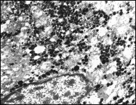
Anti-Goat IgG (whole molecule)-Gold antibody is suitable for use in immunoelectron microscopy. The antibody can also be used for dot blot.
Physical form
Colloidal suspension in Tris buffered saline, pH 8.2, with 30% glycerol (v/v), 1% bovine serum albumin (w/v) and 15 mM sodium azide
Disclaimer
Unless otherwise stated in our catalog or other company documentation accompanying the product(s), our products are intended for research use only and are not to be used for any other purpose, which includes but is not limited to, unauthorized commercial uses, in vitro diagnostic uses, ex vivo or in vivo therapeutic uses or any type of consumption or application to humans or animals.
Not finding the right product?
Try our Product Selector Tool.
Storage Class Code
10 - Combustible liquids
WGK
WGK 3
Flash Point(F)
Not applicable
Flash Point(C)
Not applicable
Personal Protective Equipment
dust mask type N95 (US), Eyeshields, Gloves
Choose from one of the most recent versions:
Already Own This Product?
Find documentation for the products that you have recently purchased in the Document Library.
Customers Also Viewed
Characterization of Immunological Interactions at an Immunoelectrode by Scanning Electron Microscopy
Ambrosi, A., et al.
Electroanalysis, 19(2-3), 244-252 (2007)
Shujiang Shang et al.
The Journal of cell biology, 215(3), 369-381 (2016-11-02)
Transient receptor potential A1 (TRPA1) is a nonselective cation channel implicated in thermosensation and inflammatory pain. In this study, we show that TRPA1 (activated by allyl isothiocyanate, acrolein, and 4-hydroxynonenal) elevates the intracellular Ca2+ concentration ([Ca2+]i) in dorsal root ganglion
N A Bright et al.
Journal of anatomy, 184 ( Pt 2), 297-308 (1994-04-01)
The human fetus acquires maternal IgG via the chorioallantoic placenta. Utilising antibodies against 3 characterised subtypes of IgG receptor (Fc gamma R) expressed by human leucocytes, we show by confocal immunofluorescence microscopy that these molecules are also expressed by cells
L Moysset et al.
Planta, 213(4), 565-574 (2001-09-15)
The intracellular localization of phytochrome in the pulvini of Robinia pseudoacacia L. was analyzed by immunogold electron microscopy after red (R; 15 min) and far-red (FR; 5 min) irradiation 2 h after the beginning of the photoperiod. Screening of the
Elie Akoury et al.
Human reproduction (Oxford, England), 30(1), 159-169 (2014-11-02)
What is the subcellular localization in human oocytes and preimplantation embryos, of the two maternal-effect proteins, NLRP7 and KHDC3L, responsible for recurrent hydatidiform moles (RHMs)? NLRP7 and KHDC3L localize to the oocyte cytoskeleton and are polar and absent from the
Our team of scientists has experience in all areas of research including Life Science, Material Science, Chemical Synthesis, Chromatography, Analytical and many others.
Contact Technical Service


