Skip to Content
MilliporeSigma
All Photos(1)

Documents

MABT205

Sigma-Aldrich

Anti-α-Tubulin Antibody, clone DM1A

clone DM1A, from mouse

Synonym(s):

Tubulin alpha-1 chain

Sign Into View Organizational & Contract Pricing


About This Item

UNSPSC Code:
12352203
eCl@ss:
32160702
NACRES:
NA.41

biological source

mouse

Quality Level

antibody form

purified immunoglobulin

antibody product type

primary antibodies

clone

DM1A, monoclonal

species reactivity

mouse, human

species reactivity (predicted by homology)

chicken (based on 100% sequence homology)

technique(s)

immunocytochemistry: suitable
immunohistochemistry: suitable
western blot: suitable

isotype

IgG1κ

UniProt accession no.

shipped in

wet ice

target post-translational modification

unmodified

Gene Information

human ... TUBA1A(7846)

General description

Tubulin is one of the main components of the cytoskeleton. It is a heterodimeric structure consisting of interlocking alpha and beta chains. Like other cytoskeleton proteins, tubulin undergoes dynamic changes depending on the cellular context, which are mediated by binding and release of GDP/GTP. Tubulin proteins may also undergo post-translational modifications, and are essential to cellular transport and mitotic processes.

Immunogen

Native chicken brain microtubules.

Application

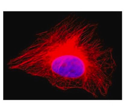
Immunocytochemistry Analysis: A 1:500 dilution from a representative lot detected α-tubulin in NIH/3T3, A431, and HeLa cells.

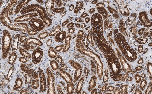
Immunohistochemistry Analysis: A 1:10,000 dilution from a representative lot detected α-tubulin in normal human brain tissue, human brain cancer tissue, normal rat hippocampus tissue and normal rat cerebellum tissue.
Research Category
Cell Structure
Research Sub Category
Cytoskeleton
This Anti-α-Tubulin Antibody, clone DM1A is validated for use in Western Blotting, ICC, IHC for the detection of α-Tubulin.

Quality

Evaluated by Western Blot in HeLa cell lysate.

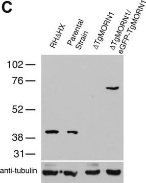
Western Blot Analysis: 0.01 µg/mL of this antibody detected α-tubulin in 10 µg of HeLa cell lysate.

Target description

~60 kDa observed. Uniprot describes a molecular weight at ~46 kDA, but can be higher due to post-translational modification. This protein has been observed at ~55 kDa in some cell lysates (Saugstad, J. A., et al. (2002). J Neurochem. 80(6):980-988.).

Linkage

Replaces: 04-1117

Physical form

Format: Purified
Protein G Purified
Purified mouse monoclonal IgG1κ in buffer containing 0.1 M Tris-Glycine (pH 7.4), 150 mM NaCl with 0.05% sodium azide.

Storage and Stability

Stable for 1 year at 2-8°C from date of receipt.

Analysis Note

Control
HeLa cell lysate.

Other Notes

Concentration: Please refer to the Certificate of Analysis for the lot-specific concentration.

Disclaimer

Unless otherwise stated in our catalog or other company documentation accompanying the product(s), our products are intended for research use only and are not to be used for any other purpose, which includes but is not limited to, unauthorized commercial uses, in vitro diagnostic uses, ex vivo or in vivo therapeutic uses or any type of consumption or application to humans or animals.

Not finding the right product?  

Try our Product Selector Tool.

Storage Class Code

12 - Non Combustible Liquids

WGK

WGK 1

Flash Point(F)

Not applicable

Flash Point(C)

Not applicable


Certificates of Analysis (COA)

Search for Certificates of Analysis (COA) by entering the products Lot/Batch Number. Lot and Batch Numbers can be found on a product’s label following the words ‘Lot’ or ‘Batch’.

Already Own This Product?

Find documentation for the products that you have recently purchased in the Document Library.

Visit the Document Library

Shanshan Zhao et al.
Experimental & molecular medicine, 51(7), 84-84 (2019-07-25)
Focal cortical dysplasia type II (FCDII) is a cerebral cortex malformation characterized by local cortical structure disorganization, neuronal dysmorphology, and refractory epilepsy. Brain somatic mutations in several genes involved in the PI3K/AKT/mTOR pathway are associated with FCDII, but they are
Noelia Prieto-Martínez et al.
Reproduction, fertility, and development, 28(6), 663-672 (2014-12-09)
Aquaporins (AQPs) are integral membrane water channels that allow transport of water and small solutes across cell membranes. Although water permeability is known to play a critical role in mammalian cells, including spermatozoa, little is known about their localisation in
Yiru Jiang et al.
Molecular therapy. Methods & clinical development, 27, 246-258 (2022-11-03)
Fragile X syndrome (FXS) is caused by the loss of the fragile X messenger ribonucleoprotein 1 (FMRP) encoded by the FMR1 gene. Gene therapy using adeno-associated virus (AAV) to restore FMRP expression is a promising therapeutic strategy. However, so far
Laurent Jullien et al.
Nucleic acids research, 41(2), 900-911 (2012-11-30)
Telomeres cap the ends of chromosomes and regulate the replicative life span of human somatic cells. Telomere function is lost upon critical shortening and a p53-dependent checkpoint that detects altered telomere states at the G1/S transition was proposed to act
Trevor M Morey et al.
The Journal of biological chemistry, 290(18), 11443-11454 (2015-04-02)
Hyperthermia is a proteotoxic stress that is lethal when exposure is extreme but also cytoprotective in that sublethal exposure leads to the synthesis of heat shock proteins, including HSP70, which are able to inhibit stress-induced apoptosis. CDK5 is an atypical

Our team of scientists has experience in all areas of research including Life Science, Material Science, Chemical Synthesis, Chromatography, Analytical and many others.

Contact Technical Service