Skip to Content
MilliporeSigma
All Photos(2)

Key Documents

MABN1801

Sigma-Aldrich

Anti-PMCA4b Antibody, clone JA3

clone JA3, from mouse

Synonym(s):

Plasma membrane calcium-transporting ATPase 4, EC: 3.6.3.8, Matrix-remodeling-associated protein 1, Plasma membrane calcium ATPase isoform 4, Plasma membrane calcium pump isoform 4

Sign Into View Organizational & Contract Pricing


About This Item

UNSPSC Code:
12352203
eCl@ss:
32160702
NACRES:
NA.41

biological source

mouse

Quality Level

antibody form

purified immunoglobulin

antibody product type

primary antibodies

clone

JA3, monoclonal

species reactivity

chimpanzee, human

packaging

antibody small pack of 25 μg

technique(s)

ELISA: suitable
immunocytochemistry: suitable
immunohistochemistry: suitable (paraffin)
western blot: suitable

isotype

IgG1κ

NCBI accession no.

UniProt accession no.

shipped in

ambient

target post-translational modification

unmodified

Gene Information

human ... ATP2B4(493)

General description

Plasma membrane calcium-transporting ATPase 4 (UniProt: P23634; also known as EC: 3.6.3.8, PMCA4, Matrix-remodeling-associated protein 1, Plasma membrane calcium ATPase isoform 4, Plasma membrane calcium pump isoform 4) is encoded by the ATP2B4 (also known as ATP2B2, MXRA1) gene (Gene ID: 493) in human. PMCA4 is a multi-pass membrane protein that acts as a calcium/calmodulin-regulated and magnesium-dependent enzyme and catalyzes the hydrolysis of ATP coupled with the transport of calcium out of the cell. PMCA4 contains 2 calmodulin binding regions known as subdomain A (aa 1086-1103) and B (aa 1104-1113) and a PDZ domain-binding motif that lies within the last 7 amino acids of the carboxy terminus, this latter is absent in the PMCAa tail. PMCA4 is shown to be a regulator of neuronal nitric-oxide synthase (nNOS) and is suggested to play a structural role in tethering nNOS to a highly compartmentalized domain in the cardiac cell membrane. PMCA4b is quite ubiquitousi is a major PMCA expressed in developing mammary tissue. The kinetics of binding of Ca2+/CaM to PMCA4b is very slow, but remains active for longer periods when compared to PMCA4a. A significantly reduced migration of PCMA4b expressing in B-RAF mutant cells is linked to their reduced metastatic potential. (Ref.: Strehler, EE. (2015). Biochem. Biophys. Res. Commun. 460(1); 26-33; Afronze, T., et al. (2014). J. Biol. Chem.289 (10), 7221-31); Mohanmed TM et al. (2011). J. Biol. Chem. 286(48):41520-29).

Specificity

Clone JA3 detects Plasma membrane calcium-transporting ATPase in human brain tissue. It targets an epitope with in 25 amino acids from the C-terminal region.

Immunogen

Purified plasma membrane calcium-transporting ATPase from human red blood cells.

Application

Anti-PMCA4b, clone JA3, Cat. No. MABN1801, is a highly specific mouse monoclonal antibody that targets PMCA4b and has been tested in ELISA, Immunocytochemistry, Immunohistochemistry (Paraffin), and Western Blotting.
Research Category
Neuroscience
Western Blotting Analysis: A representative lot detected PMCA4b in Western Blotting applications (Caride, A.J., et. al. (1996). Biochem J. 316 (Pt 1):353-9; Borke, J.L., et. al. (1987). J clin Invest. 80(5):1225-31; DeMarco, S.J., et. al. (2002). J Biol Chem. 277(12):10506-11; Gros, R., et. al. (2003). Circ Res. 93(7):614-21; Paszty, K., et. al. (2015). Sci Signal. 8(364):ra19; Usachev. M., et. al. (2001). J Neurochem. 76(6):1756-65).

Immunohistochemistry Analysis: A 1:50-250 dilution from a representative lot detected PMCA4b in human cerebral cortex, human kidney, and bovine cornea tissues.

Immunohistochemistry Analysis: A representative lot detected PMCA4b in Immunohistochemistry applications (Borke, J.L., et. al. (1987). J clin Invest. 80(5):1225-31).

Immunocytochemistry Analysis: A representative lot detected PMCA4b in Immunocytochemistry applications (Usachev. M., et. al. (2002). J Neuron. 33(1):113-22).

ELISA Analysis: A representative lot detected PMCA4b in ELISA applications (Caride, A.J., et. al. (1996). Biochem J. 316 (Pt 1):353-9; Borke, J.L., et. al. (1987). J clin Invest. 80(5):1225-31).

Quality

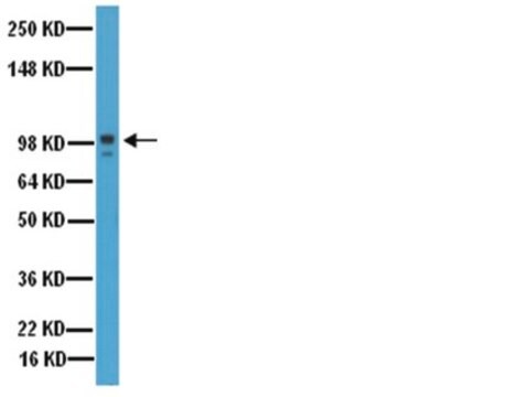
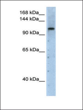
Evaluated by Western Blotting in human brain tissue lysate.

Western Blotting Analysis: 0.5 µg/mL of this antibody detected PMCA4b in 10 µg of human brain tissue lysate.

Target description

~134 kDa observed; 137.92 kDa calculated. Uncharacterized bands may be observed in some lysate(s).

Physical form

Format: Purified
Protein G purified
Purified mouse monoclonal antibody IgG1 in buffer containing 0.1 M Tris-Glycine (pH 7.4), 150 mM NaCl with 0.05% sodium azide.

Storage and Stability

Stable for 1 year at 2-8°C from date of receipt.

Other Notes

Concentration: Please refer to lot specific datasheet.

Disclaimer

Unless otherwise stated in our catalog or other company documentation accompanying the product(s), our products are intended for research use only and are not to be used for any other purpose, which includes but is not limited to, unauthorized commercial uses, in vitro diagnostic uses, ex vivo or in vivo therapeutic uses or any type of consumption or application to humans or animals.

Not finding the right product?  

Try our Product Selector Tool.

Storage Class Code

12 - Non Combustible Liquids

WGK

WGK 1


Certificates of Analysis (COA)

Search for Certificates of Analysis (COA) by entering the products Lot/Batch Number. Lot and Batch Numbers can be found on a product’s label following the words ‘Lot’ or ‘Batch’.

Already Own This Product?

Find documentation for the products that you have recently purchased in the Document Library.

Visit the Document Library

Randa Naffa et al.
Cells, 9(5) (2020-05-18)
Metastatic melanoma is the most aggressive type of skin cancer. Previously, we identified the plasma membrane Ca2+ pump isoform 4b (PMCA4b or ATP2B4) as a putative metastasis suppressor in BRAF mutant melanoma cells. Metastasis suppressors are often downregulated in cancer

Our team of scientists has experience in all areas of research including Life Science, Material Science, Chemical Synthesis, Chromatography, Analytical and many others.

Contact Technical Service