Skip to Content
MilliporeSigma
All Photos(2)

Documents

MABF294

Sigma-Aldrich

Anti-LAG3 Antibody, clone 8C7.1

culture supernatant, clone 8C7.1, from mouse

Synonym(s):

Lymphocyte activation gene 3 protein, LAG-3, Protein FDC, CD223, LAG3

Sign Into View Organizational & Contract Pricing


About This Item

UNSPSC Code:
12352203
eCl@ss:
32160702
NACRES:
NA.41

biological source

mouse

Quality Level

antibody form

culture supernatant

antibody product type

primary antibodies

clone

8C7.1, monoclonal

species reactivity

human

technique(s)

flow cytometry: suitable
western blot: suitable

isotype

IgG2aκ

NCBI accession no.

UniProt accession no.

shipped in

dry ice

target post-translational modification

unmodified

Gene Information

human ... LAG3(3902)

General description

Lymphocyte activation gene 3 protein (UniProt P18627; also known as CD223, LAG-3, Protein FDC) is encoded by the LAG3 (also known as FDC) gene (Gene ID 3902) in human. LAG3 is a single-pass type I membrane protein that binds MHC class II molecules on antigen presenting cells with high affinity. LAG3 expression is induced after T cell activation and functions as a negative regulator of T cell function. LAG3 blockage is shown to augment T cell proliferation and cytokine production, and LAG3-deficient mice have a defect in the downregulation of T cell responses induced by staphylococcal enterotoxin B or by virus infection. In addition, LAG3 is expressed on activated regulatory T (Treg) cells at higher levels than on activated effector T cells. LAG3 blockage inhibits the suppressive activity of Treg cells. LAG3 is also upregulated on tumour-infiltrating lymphocytes (TILs), and LAG3 blockage can enhance anti-tumour T cell responses.

Immunogen

Mouse Fc-tagged recombinant human LAG3 extracellular fragment.

Application

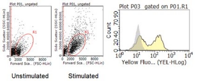
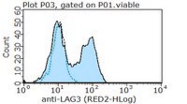
Flow Cytometry Analysis: A 1:100 dilution of this antibody from a representative lot detected LAG3 in 1X10E6 Jurkat cells subjected to CD3/CD28 costimulation.
This Anti-LAG3 Antibody, clone 8C7.1 is validated for use in Western Blotting, Flow Cytometry for the detection of LAG3.

Quality

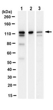
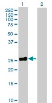
Evaluated by Western Blotting in NK-92c ell lysate.

Western Blotting Analysis: A 1:1,000 dilution of this antibody detected LAG3 in 10 µg of NK-92 cell lysate.

Target description

~60 kDa observed

Physical form

Mouse monoclonal IgG2aκ antibody tissue culture supernatant with 0.05% sodium azide.

Other Notes

Concentration: Please refer to lot specific datasheet.

Not finding the right product?  

Try our Product Selector Tool.

Storage Class Code

10 - Combustible liquids

WGK

WGK 2


Certificates of Analysis (COA)

Search for Certificates of Analysis (COA) by entering the products Lot/Batch Number. Lot and Batch Numbers can be found on a product’s label following the words ‘Lot’ or ‘Batch’.

Already Own This Product?

Find documentation for the products that you have recently purchased in the Document Library.

Visit the Document Library

Our team of scientists has experience in all areas of research including Life Science, Material Science, Chemical Synthesis, Chromatography, Analytical and many others.

Contact Technical Service