Skip to Content
MilliporeSigma
All Photos(1)

Key Documents

MAB3747

Sigma-Aldrich

Anti-Microphthalmia (Mi) Antibody, clone C5

clone C5, Chemicon®, from mouse

Sign Into View Organizational & Contract Pricing


About This Item

UNSPSC Code:
12352203
eCl@ss:
32160702
NACRES:
NA.41

biological source

mouse

Quality Level

antibody form

purified immunoglobulin

antibody product type

primary antibodies

clone

C5, monoclonal

species reactivity

mouse

species reactivity (predicted by homology)

human, rat

manufacturer/tradename

Chemicon®

technique(s)

electrophoretic mobility shift assay: suitable
immunohistochemistry (formalin-fixed, paraffin-embedded sections): suitable
immunoprecipitation (IP): suitable
western blot: suitable

isotype

IgG1

shipped in

wet ice

target post-translational modification

unmodified

General description

MiTF (Microphthalmia associated transcription factor) is a basic helix loop helix leucine zipper (b HLH ZIP) transcription factor implicated in pigmentation, mast cells and bone development. Mutations in MiTF cause auditory pigmentary syndromes, such as Waardenburg syndrome type II, type IIa and Tietz syndrome in humans. There are two known isoforms of MiTF differing by 66 amino acids at the NH2 terminus. Shorter forms are expressed in melanocytes and run as two bands at 52 kDa and 56 kDa, while the longer Mi form runs as a cluster of bands at 60-70 kDa in osteoclasts and in B16 melonoma cells (but not other melanoma cell lines), as well as mast cells and heart. MiTF plays a critical role in the differentiation of various cell types as neural crest-derived melanocytes, mast cells, osteoclasts and optic cup-derived retinal pigment epithelium. Mi is a basic helix-loop-helix-leucin zipper (b-HLH-ZIP) transtripotion factor implicated in pigmentation, mast cells and bone development. The mutation of Mi causes Waardenburg Syndrome type II in humans. In mice, a profound loss of pigmented cells in the skin eye and inner ear results, as well as osteopetrosis and defects in natural killer and mast cells. These melanocyte isoforms have been shown by two dimensional tryptic mapping to differ in c-Kit-induced phosphorylation. Osteopetrotic rat strain harbors a large genomic deletion encompassing the 3′ half of Mi including most of the b-HLH-ZIP region. Osteoclasts from these animals lack Mi protein in contrast to wild-type rat, mouse, and human osteoclasts.

Specificity

In Western blotting, it recognizes a doublet of 52-56kDa, identified as serine-phosphorylated and unphosphorylated forms of melanocytic isoforms of microphthalmia (Mi). There are two known isoforms of Mi differing by 66 amino acids at the NH2 terminus. Shorter forms are expressed in melanocytes and run as two bands at 52kDa and 56kDa, while the longer Mi form runs as a cluster of bands at 60-70kDa in osteoclasts and in B16 melonoma cells (but not other melanoma cell lines), as well as mast cells andheart. It reacts with both melanocytic as well as the nonmelanocytic isoforms of Mi. This Ab does not cross-react with other b-HLH-ZIP factors by DNA mobility shift assay.

Immunogen

Hybridoma produced by the fusion of splenocytes from RBF/DnJ mice immunized with an N-terminal fragment of human microphthalmia protein and mouse myeloma NS1 cells.

Application

Anti-Microphthalmia (Mi) Antibody, clone C5 is a Mouse Monoclonal Antibody for detection of Microphthalmia (Mi) also known as Microphthalmia-associated transcription factor & has been tested in EMSA, IHC, IHC(P), IP & WB.
Immunoprecipitation:
A previous lot of this antibody was used in IP. (2 µg/mg of protein lysate)

Gel supershift assays:
A previous lot of this antibody was used in EMSA.

Immunohistochemistry(paraffin):
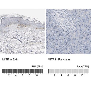
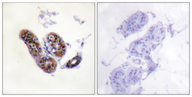
A previous lot of this antibody was used in IH on frozen and formalin/paraffin tissue sections.

Optimal working dilutions must be determined by end user.

Quality

Routinely evaluated by Western Blot on Mouse Brain lysates.

Western Blot Analysis:
1:500 dilution of this lot detected Mi on 10 μg of Mouse Brain lysates.

Target description

53-56 kDa

Physical form

Format: Purified
Purified mouse monoclonal IgG1 in phosphate buffered saline with 0.08% sodium azide.

Storage and Stability

Stable for 1 year at -20ºC in undiluted aliquots from date of receipt.
Handling Recommendations: Upon receipt, and prior to removing the cap, centrifuge the vial and gently mix the solution. Aliquot into microcentrifuge tubes and store at -20°C. Avoid repeated freeze/thaw cycles, which may damage the IgG1 and affect product performance.

Analysis Note

Control
501 Mel human melanoma cells, wild-type human, rat, mouse osteoclast cells.

Other Notes

Concentration: Please refer to the Certificate of Analysis for the lot-specific concentration.

Legal Information

CHEMICON is a registered trademark of Merck KGaA, Darmstadt, Germany

Not finding the right product?  

Try our Product Selector Tool.

Storage Class Code

12 - Non Combustible Liquids

WGK

WGK 2

Flash Point(F)

Not applicable

Flash Point(C)

Not applicable


Certificates of Analysis (COA)

Search for Certificates of Analysis (COA) by entering the products Lot/Batch Number. Lot and Batch Numbers can be found on a product’s label following the words ‘Lot’ or ‘Batch’.

Already Own This Product?

Find documentation for the products that you have recently purchased in the Document Library.

Visit the Document Library

Inhibitory effects of 1-O-methyl-fructofuranose from Schisandra chinensis fruit on melanogenesis in B16F0 melanoma cells.
Eun Young Oh,Ji Yeon Jang,Yung Hyun Choi,Young Whan Choi,Byung Tae Choi
Journal of Ethnopharmacology null
T J Hemesath et al.
Nature, 391(6664), 298-301 (1998-01-24)
Germline mutations at loci encoding the transcription factor Microphthalmia (Mi), the cytokine receptor c-Kit, or its ligand Steel factor (S1) result in strikingly similar defects in mast cell and melanocyte development. Here we describe a biochemical link between Kit signalling
Ji Yeon Jang et al.
Journal of ethnopharmacology, 137(3), 1207-1214 (2011-08-06)
Ethnopharmacological relevance Nardostachys chinensis has been used in folk medicine to treat melasma and lentigines in Korea. We investigated the inhibitory activities of Nardostachys chinensis in melanogenesis and its related signaling pathway. Bioassay-guided fractionation of Nardostachys chinensis using solvent partitioning
Pakavarin Louphrasitthiphol et al.
Molecular cell, 79(3), 472-487 (2020-06-13)
It is widely assumed that decreasing transcription factor DNA-binding affinity reduces transcription initiation by diminishing occupancy of sequence-specific regulatory elements. However, in vivo transcription factors find their binding sites while confronted with a large excess of low-affinity degenerate motifs. Here, using
Chelsey D Kline et al.
Cancers, 14(20) (2022-10-28)
TR1 and other selenoproteins have paradoxical effects in melanocytes and melanomas. Increasing selenoprotein activity with supplemental selenium in a mouse model of UV-induced melanoma prevents oxidative damage to melanocytes and delays melanoma tumor formation. However, TR1 itself is positively associated

Our team of scientists has experience in all areas of research including Life Science, Material Science, Chemical Synthesis, Chromatography, Analytical and many others.

Contact Technical Service