Skip to Content
MilliporeSigma
All Photos(2)

Key Documents

MAB1926-I

Sigma-Aldrich

Anti-Fibronectin Antibody, cell binding domain Antibody, clone P1H11

clone P1H11, from mouse

Synonym(s):

FN, Cold-insoluble globulin, CIG

Sign Into View Organizational & Contract Pricing


About This Item

UNSPSC Code:
12352203
eCl@ss:
32160702
NACRES:
NA.41

biological source

mouse

Quality Level

antibody form

purified antibody

antibody product type

primary antibodies

clone

P1H11, monoclonal

species reactivity

human, porcine, mouse

packaging

antibody small pack of 25 μL

technique(s)

immunocytochemistry: suitable
immunohistochemistry: suitable (paraffin)
western blot: suitable

isotype

IgG1κ

NCBI accession no.

UniProt accession no.

shipped in

ambient

target post-translational modification

unmodified

Gene Information

human ... FN1(2335)
mouse ... Fn1(14268)

General description

Fibronectin (UniProt: P02751; also known as FN, Cold-insoluble globulin, CIG) is encoded by the FN1 (also known as FN) gene (Gene ID: 2335) in human. Fibronectin is are involved various biological activities, including cell adhesion, cell motility, opsonization, wound healing, and maintenance of cell shape. It is essential for osteoblast mineralization and participates in the regulation of type I collagen deposition by osteoblasts. Fibronectin is usually present as homodimer of ~250 kDa subunits where subunits are connected by two disulfide bonds at the C-terminal region. Each monomer contains three repeating units, which are designated as type I, II, and III repeats containing about 40, 60, and 90 amino acids, respectively. Plasma fibronectin (soluble dimeric form) is secreted by hepatocytes and the cellular fibronectin (dimeric or cross-linked multimeric forms) is produced by fibroblasts, epithelial and other cell types. Cellular fibronectin is deposited as fibrils in the extracellular matrix. Fibronectin is recognized by integrins alpha5beta1 and alphaVbeta3. The primary sequence motif of fibronectin for integrin binding is a tripeptide, Arg-Gly-Asp (RGD). Mutations in FN gene are known to cause glomerulopathy that is characterized by proteinuria, microscopic hematuria, and hypertension, which can lead to end-stage renal failure.

Specificity

Clone P1H11 detects fibronectin in human.

Immunogen

Human Plasma Fibronectin.

Application

Anti-Fibronectin, cell binding domain, clone P1H11, Cat. No. MAB1926-I is a mouse monoclonal antibody that detects human fibronectin and has been tested for use in Immunocytochemistry, Immunohistochemistry (Paraffin), and Western Blotting.
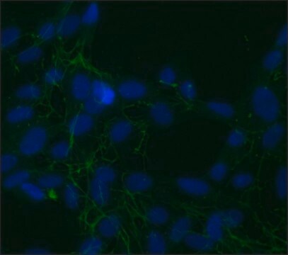
Immunohistochemistry Analysis: A 1:50 dilution from a representative lot detected Fibronectin in human kidney and human cerebellum tissues.

Western Blotting Analysis: A representative lot detected Fibronectin in Western Blotting applications (Zhang, H., et. al. (2010). Am J Physiol Renal Physiol. 299(1):F91-8).

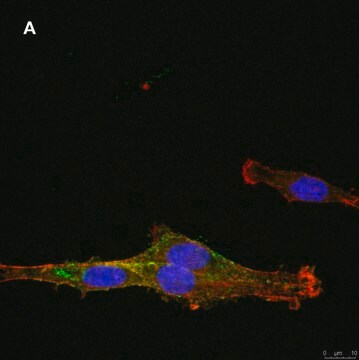
Immunocytochemistry Analysis: A representative lot detected Fibronectin in Immunocytochemistry applications (Mardilovich, A., et. al. (2006). Langmuir. 22(7):3259-64).

Immunocytochemistry Analysis: A representative lot detected Fibronectin in Immunocytochemistry applications (Hofanson, D.M., et. al. (2010). Biomaterials. 31(26):6730-7).

Immunocytochemistry Analysis: A representative lot detected Fibronectin in Immunocytochemistry applications (Waterman, R.S., et. al. (2010). PLoS One. 5(4):e10088).

Immunohistochemistry Analysis: A representative lot detected Fibronectin in Immunohistochemistry applications (Carlos, C.P., et. al. (2014). PLoS One. 9(7):e103660).

Western Blotting Analysis: A representative lot detected Fibronectin in Western Blotting applications (Zhang, H., et. al. (2008). Am J Physiol Renal Physiol. 295(4):F1071-81).
Research Category
Cell Structure

Quality

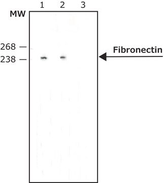
Evaluated by Western Blotting in human thymus tissue lysate.

Western Blotting Analysis: 1 µg/mL of this antibody detected Fibronectin in 10 µg of human thymus tissue lysate.

Target description

~270 kDa observed; 262.62 kDa calculated. Uncharacterized bands may be observed in some lysate(s).

Physical form

Format: Purified
Protein G purified
Purified mouse monoclonal antibody IgG1 in buffer containing 0.1 M Tris-Glycine (pH 7.4), 150 mM NaCl with 0.05% sodium azide.

Storage and Stability

Stable for 1 year at 2-8°C from date of receipt.

Other Notes

Concentration: Please refer to lot specific datasheet.

Disclaimer

Unless otherwise stated in our catalog or other company documentation accompanying the product(s), our products are intended for research use only and are not to be used for any other purpose, which includes but is not limited to, unauthorized commercial uses, in vitro diagnostic uses, ex vivo or in vivo therapeutic uses or any type of consumption or application to humans or animals.

Not finding the right product?  

Try our Product Selector Tool.

Storage Class Code

12 - Non Combustible Liquids

WGK

WGK 1

Flash Point(F)

Not applicable

Flash Point(C)

Not applicable


Certificates of Analysis (COA)

Search for Certificates of Analysis (COA) by entering the products Lot/Batch Number. Lot and Batch Numbers can be found on a product’s label following the words ‘Lot’ or ‘Batch’.

Already Own This Product?

Find documentation for the products that you have recently purchased in the Document Library.

Visit the Document Library

Shirley Genah et al.
Biomedicines, 9(3) (2021-04-04)
The fine control of inflammation following injury avoids fibrotic scars or impaired wounds. Due to side effects by anti-inflammatory drugs, the research is continuously active to define alternative therapies. Among them, physical countermeasures such as photobiomodulation therapy (PBMT) are considered

Our team of scientists has experience in all areas of research including Life Science, Material Science, Chemical Synthesis, Chromatography, Analytical and many others.

Contact Technical Service