Skip to Content
MilliporeSigma
All Photos(1)

Key Documents

AB16503

Sigma-Aldrich

Anti-Apaf-1 Antibody, CT

Chemicon®, from rabbit

Synonym(s):

Apoptosis Protease-activating Factor 1

Sign Into View Organizational & Contract Pricing


About This Item

UNSPSC Code:
12352203
eCl@ss:
32160702
NACRES:
NA.41

biological source

rabbit

Quality Level

antibody form

affinity purified immunoglobulin

antibody product type

primary antibodies

clone

polyclonal

species reactivity

human, rat, mouse

manufacturer/tradename

Chemicon®

technique(s)

western blot: suitable

UniProt accession no.

shipped in

wet ice

target post-translational modification

unmodified

Gene Information

human ... APAF1(317)

Specificity

Apoptosis is related to many diseases and is induced by a family of cell death receptors and their ligands. Cell death signals are transduced by death domain containing adapter molecules and members of the caspase family of proteases. The mammalian homologues of the key cell death gene CED-4 in C. elegans has been identified recently from human and mouse and was designated Apaf1 (for apoptosis protease-activating factor 1; Zou et al. 1997; Cecconi et al. 1998). Apaf1 binds to cytochrome C (Apaf2) and caspase-9 (Apaf3), which leads to caspase-9 activation. Activated caspase-9 in turn cleaves and activates caspase-3 which is one of the key proteases, being responsible for the proteolytic cleavage of many key proteins in apoptosis (Li et al. 1997). Apaf1 can also associate with caspase-4 and caspase-8 (Hu et al. 1998). Apaf1 is ubiquitously expressed in human tissues (Zou et al. 1997).

Immunogen

Epitope: C-terminus
Rabbit anti-Apaf1 (C-terminus) polyclonal antibody was raised against a peptide corresponding to amino acids 1158 to 1177 of human Apaf1 (Zou et al. 1997). The sequence of the immunogenic peptide differs from that of murine Apaf1 by one amino acid (Zou et al. 1997; Cecconi et al. 1998).

Application

Anti-Apaf-1 Antibody, C-terminus detects level of Apaf-1 & has been published & validated for use in WB.
Research Category
Apoptosis & Cancer
Research Sub Category
Apoptosis - Additional
Western blot: 1:500 to 1:2000.

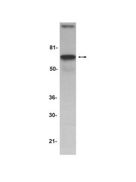
Human heart tissue lysate can be used as positive control and a 130 kDa band should be detected.

Optimal working dilutions must be determined by end user.

Linkage

Replaces: 04-579

Physical form

Format: Purified
Purified IgG by immunoaffinity chromatography. Liquid in PBS containing 0.02% sodium azide.

Storage and Stability

Maintain refrigerated at 2-8°C in undiluted aliquots for up to 12 months.

Other Notes

Concentration: Please refer to the Certificate of Analysis for the lot-specific concentration.

Legal Information

CHEMICON is a registered trademark of Merck KGaA, Darmstadt, Germany

Disclaimer

Unless otherwise stated in our catalog or other company documentation accompanying the product(s), our products are intended for research use only and are not to be used for any other purpose, which includes but is not limited to, unauthorized commercial uses, in vitro diagnostic uses, ex vivo or in vivo therapeutic uses or any type of consumption or application to humans or animals.

Not finding the right product?  

Try our Product Selector Tool.

Storage Class Code

10 - Combustible liquids

WGK

WGK 2

Flash Point(F)

Not applicable

Flash Point(C)

Not applicable


Certificates of Analysis (COA)

Search for Certificates of Analysis (COA) by entering the products Lot/Batch Number. Lot and Batch Numbers can be found on a product’s label following the words ‘Lot’ or ‘Batch’.

Already Own This Product?

Find documentation for the products that you have recently purchased in the Document Library.

Visit the Document Library

Age-related alterations in expression of apoptosis regulatory proteins and heat shock proteins in rat skeletal muscle.
Linda Chung, Yuk-Chow Ng
Biochimica et Biophysica Acta null
Ignacio Vega-Naredo et al.
Methods in molecular biology (Clifton, N.J.), 2310, 161-178 (2021-06-08)
Mitochondria play a key role in cell death and its regulation. The permeabilization of the outer mitochondrial membrane, which is mainly controlled by proteins of the BCL-2 family, is a key event that can be directly induced by different signaling
Parco M Siu et al.
American journal of physiology. Cell physiology, 288(2), C338-C349 (2004-10-16)
The influence of aging on skeletal myocyte apoptosis is not well understood. In this study we examined apoptosis and apoptotic regulatory factor responses to muscle atrophy induced via limb unloading following loading-induced hypertrophy. Muscle hypertrophy was induced by attaching a
F Cecconi et al.
Cell, 94(6), 727-737 (1998-09-30)
The cytosolic protein APAF1, human homolog of C. elegans CED-4, participates in the CASPASE 9 (CASP9)-dependent activation of CASP3 in the general apoptotic pathway. We have generated by gene trap a null allele of the murine Apaf1. Homozygous mutants die
Parco M Siu et al.
FASEB journal : official publication of the Federation of American Societies for Experimental Biology, 18(10), 1150-1152 (2004-05-11)
The effect of exercise on apoptosis in postmitotic tissues is not known. In this study, we investigated the effect of regular moderate physical activity (i.e., exercise training) on the extent of apoptosis in rat skeletal and cardiac muscles. Adult Sprague

Our team of scientists has experience in all areas of research including Life Science, Material Science, Chemical Synthesis, Chromatography, Analytical and many others.

Contact Technical Service