Skip to Content
MilliporeSigma
All Photos(7)

Key Documents

AB1508

Sigma-Aldrich

Anti-Glutamate Receptor 4 Antibody

Chemicon®, from rabbit

Synonym(s):

AMPA-selective glutamate receptor 4, Glutamate receptor ionotropic, AMPA 4, glutamate receptor 4, glutamate receptor, ionotrophic, AMPA 4

Sign Into View Organizational & Contract Pricing


About This Item

UNSPSC Code:
12352203
eCl@ss:
32160702
NACRES:
NA.41

biological source

rabbit

Quality Level

antibody form

affinity isolated antibody

antibody product type

primary antibodies

clone

polyclonal

purified by

affinity chromatography

species reactivity

mouse, rat

manufacturer/tradename

Chemicon®

technique(s)

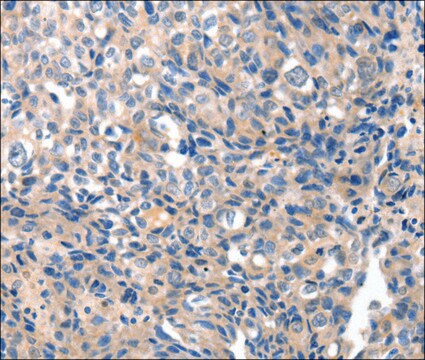
immunocytochemistry: suitable
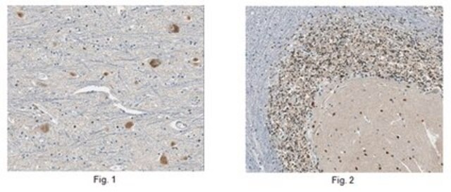
immunohistochemistry (formalin-fixed, paraffin-embedded sections): suitable
immunoprecipitation (IP): suitable
western blot: suitable

UniProt accession no.

shipped in

wet ice

target post-translational modification

unmodified

Gene Information

human ... GRIA4(2893)

General description

Glutamate receptor 4 (UniProt: P48058; also known as GluR-4, GluR4, AMPA-selective glutamate receptor 4, GluR-D, Glutamate receptor ionotropic AMPA 4, GluA4) is encoded by the GRIA4 (also known as GLUR4) gene (Gene ID: 2893) in human. The postsynaptic actions of glutamate are mediated by a variety of receptors that are named according to their selective agonists. GluR-4 is a multi-pass membrane protein that functions as ligand-gated ion channel in the central nervous system and plays an important role in excitatory synaptic transmission. It displays greater affinity for AMPA. GluR-4 is synthesized with a signal peptide (aa 1-20), which is cleaved off in the mature form. GluR-4 can exist as a homotetramer or heterotetramer of pore-forming glutamate receptor subunits, tetramers can also form by the dimerization of dimers. GluR-4 is generally palmitoylated and becomes de-palmitoylated upon glutamate stimulation. Palmitoylation on Cys 611 leads to its Golgi retention and reduced cell surface expression. However, palmitoylation at Cys 837 does not affect cell surface expression, but can regulate stimulation-dependent endocytosis. GluR-4 can undergo phosphorylation at Serine 862 by PKC gamma and this phosphorylation increases plasma membrane-associated GRI4 expression.

Specificity

Cat. # AB1508 recognizes C-Terminus of GluR4.
The immunogen sequence shares 100% conservation with chicken, mouse, rat, oppossum, human, chimp, and monkey.

Immunogen

Epitope: C-Terminus
Synthetic linear peptide.

Application

Immunohistochemistry:
Dilutions of 1:10 – 1:500 were tested on rat brain lysate.
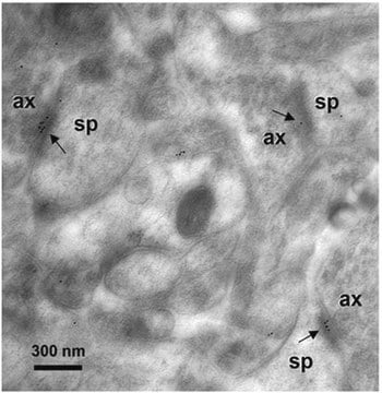
Can be used for immunocytochemistry using paraformaldehyde or paraformaldehyde/glutaraldehyde fixed tissue with light and electron microscopy. Cryostat and vibratome sections can be used with or without Triton X-100 treatment. Suggested concentrations start at 1-3 μg/mL.

Western blot analysis can be done at final concentrations of 1-3 μg/mL.

Optimal working dilutions must be determined by end user.
Research Category
Neuroscience
Research Sub Category
Neurotransmitters & Receptors
This Anti-Glutamate Receptor 4 Antibody is validated for use in IH(P), IC, IH, IP, WB for the detection of Glutamate Receptor 4.

Quality

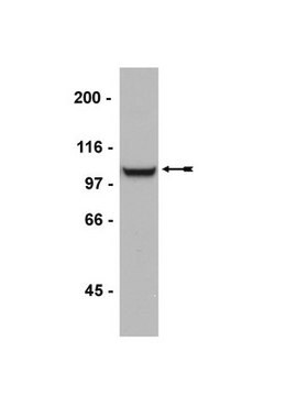
Evaluated by Western Blotting in mouse and rat brain membrane.

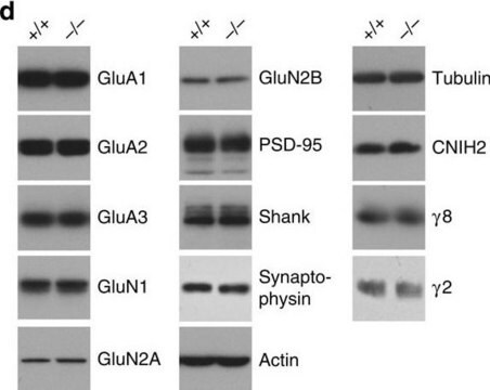
Western Blotting Analysis: A 1:500 dilution of this antibody detected Glutamate Receptor 4 in mouse and rat brain membrane.

Target description

Approx. 100 kDa

Linkage

Replaces: 06-308

Physical form

Antigen Affinity Purified
Purified rabbit polyclonal antibody in buffer containing 0.1 M Tris-Glycine (pH 7.4), 150 mM NaCl with 0.05% sodium azide.

Storage and Stability

Stable for 1 year at 2-8ºC from date of receipt.
Handling Recommendations: Upon receipt, and prior to removing the cap, centrifuge the vial and gently mix the solution. Aliquot into microcentrifuge tubes and store at -20°C.

Analysis Note

Control
Rat Brain

Other Notes

Concentration: Please refer to the Certificate of Analysis for the lot-specific concentration.

Legal Information

CHEMICON is a registered trademark of Merck KGaA, Darmstadt, Germany

Disclaimer

Unless otherwise stated in our catalog or other company documentation accompanying the product(s), our products are intended for research use only and are not to be used for any other purpose, which includes but is not limited to, unauthorized commercial uses, in vitro diagnostic uses, ex vivo or in vivo therapeutic uses or any type of consumption or application to humans or animals.

Not finding the right product?  

Try our Product Selector Tool.

Storage Class Code

12 - Non Combustible Liquids

WGK

WGK 2

Flash Point(F)

Not applicable

Flash Point(C)

Not applicable


Certificates of Analysis (COA)

Search for Certificates of Analysis (COA) by entering the products Lot/Batch Number. Lot and Batch Numbers can be found on a product’s label following the words ‘Lot’ or ‘Batch’.

Already Own This Product?

Find documentation for the products that you have recently purchased in the Document Library.

Visit the Document Library

Synaptic inputs onto small bistratified (blue-ON/yellow-OFF) ganglion cells in marmoset retina.
Percival KA, Jusuf PR, Martin PR, Grunert U
The Journal of Comparative Neurology null
Synaptic contact between melanopsin-containing retinal ganglion cells and rod bipolar cells.
?stergaard, J; Hannibal, J; Fahrenkrug, J
Investigative Ophthalmology & Visual Science null
F Yang et al.
Nature neuroscience, 4(11), 1071-1078 (2001-10-11)
Glial cell line-derived neurotrophic factor (GDNF) prevents lesion-induced death of midbrain dopaminergic neurons, but its function in normal brain remains uncertain. Here we show that GDNF acutely and reversibly potentiated the excitability of cultured midbrain neurons by inhibiting transient A-type
Cell-type-specific localization of protocadherin ?16 at AMPA and AMPAKainate receptor-containing synapses in the primate retina.
Puller C, Haverkamp S
The Journal of Comparative Neurology null
ZO-1 and the spatial organization of gap junctions and glutamate receptors in the outer plexiform layer of the mammalian retina.
Puller, C; de Sevilla Muller, LP; Janssen-Bienhold, U; Haverkamp, S
The Journal of Neuroscience null

Articles

The term neurodegeneration characterizes a chronic loss of neuronal structure and function leading to progressive mental impairments.

Our team of scientists has experience in all areas of research including Life Science, Material Science, Chemical Synthesis, Chromatography, Analytical and many others.

Contact Technical Service