Skip to Content
Merck
All Photos(1)

Key Documents

Z4751

Sigma-Aldrich

Anti-Zyxin antibody produced in rabbit

IgG fraction of antiserum, buffered aqueous solution

Synonym(s):

Anti-ESP-2, Anti-HED-2

Sign Into View Organizational & Contract Pricing


About This Item

MDL number:
UNSPSC Code:
12352203
NACRES:
NA.41

biological source

rabbit

conjugate

unconjugated

antibody form

IgG fraction of antiserum

antibody product type

primary antibodies

clone

polyclonal

form

buffered aqueous solution

mol wt

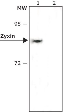
antigen 82-84 kDa

species reactivity

mouse, human, rat

technique(s)

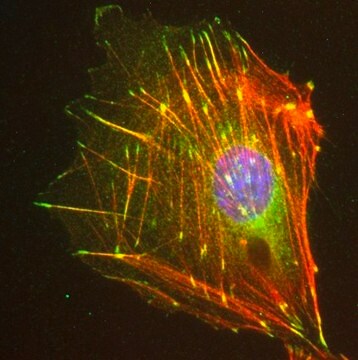
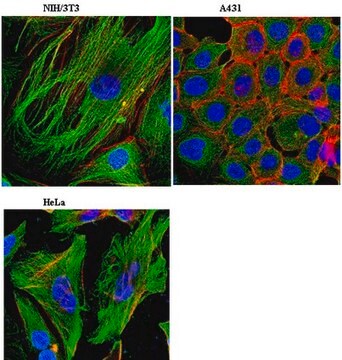
indirect immunofluorescence: 1:100 using Rat1 cells
western blot (chemiluminescent): 1:1,000 using whole extract of human HeLa cells
western blot (chemiluminescent): 1:2,000 using whole extract of mouse NIH3T3 cells

UniProt accession no.

shipped in

dry ice

storage temp.

−20°C

target post-translational modification

unmodified

Gene Information

human ... ZYX(7791)
mouse ... Zyx(22793)
rat ... Zyx(114636)

General description

Zyxin (ZYX ), a zinc-binding phosphoprotein of ∼61kDa is located at focal adhesions and along the actin cytoskeleton. It has three LIM domains. This structural protein is located on human chromosome 7q34.

Immunogen

synthetic peptide corresponding to amino acid residues 134-150 of human zyxin with N-terminal added cysteine conjugated to KLH. The corresponding sequence differs by one amino acid in mouse.

Application

Anti-Zyxin antibody has been used in fluorescence microscopy.
Applications in which this antibody has been used successfully, and the associated peer-reviewed papers, are given below.
Western Blotting (1 paper)
Applications in which this antibody has been used successfully, and the associated peer-reviewed papers, are given below.
Western Blotting (1 papers)

Biochem/physiol Actions

Zyxin (ZYX ) participates in the modulation of actin assembly at focal adhesions and stress fibers. It also regulates cell adhesion and cell cycle. Overexpression of ZYX is associated with melanoma. Suppression of ZYX  prevents cell growth.

Physical form

Solution in 0.01 M phosphate buffered saline, pH 7.4, containing 15 mM sodium azide.

Disclaimer

Unless otherwise stated in our catalog or other company documentation accompanying the product(s), our products are intended for research use only and are not to be used for any other purpose, which includes but is not limited to, unauthorized commercial uses, in vitro diagnostic uses, ex vivo or in vivo therapeutic uses or any type of consumption or application to humans or animals.

Not finding the right product?  

Try our Product Selector Tool.

Storage Class Code

10 - Combustible liquids

WGK

WGK 2

Flash Point(F)

Not applicable

Flash Point(C)

Not applicable

Personal Protective Equipment

dust mask type N95 (US), Eyeshields, Gloves

Certificates of Analysis (COA)

Search for Certificates of Analysis (COA) by entering the products Lot/Batch Number. Lot and Batch Numbers can be found on a product’s label following the words ‘Lot’ or ‘Batch’.

Already Own This Product?

Find documentation for the products that you have recently purchased in the Document Library.

Visit the Document Library

Loss of the mechanotransducer zyxin promotes a synthetic phenotype of vascular smooth muscle cells
Ghosh SK, et al.
Journal of the American Heart Association, 4(6), e001712-e001712 (2015)
Functional analysis of Zyxin in cell migration and invasive potential of oral squamous cell carcinoma cells Corrigendum in/10.3892/ijo. 2016.3702
Yamamura MN, et al.
International Journal of Oncology, 42(3), 873-880 (2013)
A cell culture model for investigation of Hirano bodies
Davis RC, et al.
Acta Neuropathologica, 115(2), 205-205 (2008)
Aniqua Rahman et al.
Molecular biology of the cell (2016-03-11)
During metastasis, cells can use proteolytic activity to form tube-like "microtracks" within the extracellular matrix (ECM). Using these microtracks, cells can migrate unimpeded through the stroma. To investigate the molecular mechanisms of microtrack migration, we developed an in vitro 3D
Richard C Davis et al.
Acta neuropathologica, 115(2), 205-217 (2007-11-06)
Hirano bodies are paracrystalline F-actin-rich aggregations associated with a variety of conditions including aging, and neurodegenerative diseases. The composition and structure of these inclusions have been described by immunohistochemistry and ultrastructure, respectively. However, studies of the physiological function and dynamics

Our team of scientists has experience in all areas of research including Life Science, Material Science, Chemical Synthesis, Chromatography, Analytical and many others.

Contact Technical Service