Skip to Content
Merck
All Photos(2)

Key Documents

A0545

Sigma-Aldrich

Anti-Rabbit IgG (whole molecule)–Peroxidase antibody produced in goat

affinity isolated antibody

Synonym(s):

Anti Rabbit, Anti Rabbit Antibody, Anti Rabbit Igg, Anti-Rabbit Igg, Goat Anti Rabbit IgG Antibody - Anti-Rabbit IgG (whole molecule)–Peroxidase antibody produced in goat, Goat Anti Rabbit Igg

Sign Into View Organizational & Contract Pricing


About This Item

MDL number:
UNSPSC Code:
12352203
NACRES:
NA.46

biological source

goat

Quality Level

conjugate

peroxidase conjugate

antibody form

affinity isolated antibody

antibody product type

secondary antibodies

clone

polyclonal

species reactivity

rabbit

should not react with

human

technique(s)

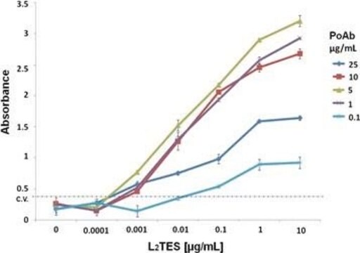
direct ELISA: 1:30,000 using using 5 μg/ml of rabbit IgG for the coating and OPD substrate
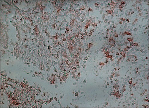
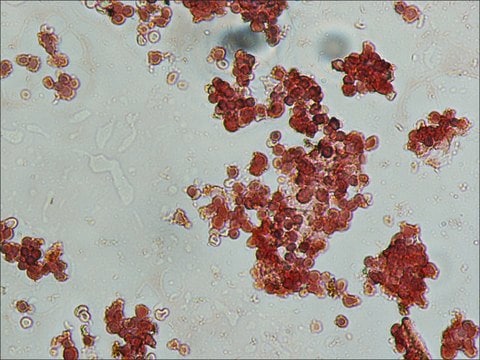
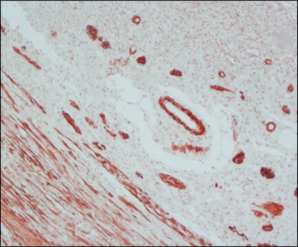
immunohistochemistry (formalin-fixed, paraffin-embedded sections): 1:200
western blot (chemiluminescent): 1:80,000-1:160,000 using detecting β-actin in total cell extract of HeLa cells (5-10 μg/mL)

shipped in

dry ice

storage temp.

−20°C

target post-translational modification

unmodified

Looking for similar products? Visit Product Comparison Guide

General description

Goat anti-rabbit IgG-peroxidase conjugate associates with all rabbit Igs. The conjugate shows no reaction with human IgG by immunoelectrophoresis (IEP). Furthermore, the antibody is adsorbed with human IgG to reduce background staining with human samples.
Immunoglobulin G (IgG) belongs to the immunoglobulin family and is a widely expressed serum antibody. An immunoglobulin has two heavy chains and two light chains connected by a disulfide bond. It is a glycoprotein and mainly helps in immune defense. IgG is a major class of immunoglobulin. Immunoglobulin G (IgG) participates in hypersensitivity type II and type III. Rabbit IgG is further divided into five classes- IgM, IgG, IgA, IgD and IgE. It has only one IgG variant with one interheavy disulfide bridge. It helps in opsonization, complement fixation and antibody dependent cell mediated cytotoxicity.

Specificity

Specificity of the Anti-Rabbit IgG-Peroxidase is determined by immunoelectrophoresis (IEP) using normal rabbit serum and rabbit IgG. The conjugate shows no reaction with human IgG by IEP.

Immunogen

Purified rabbit IgG

Application

Anti-Rabbit IgG (whole molecule)–Peroxidase antibody has been used in western blotting , immunofluorescence staining , immunochemistry and immunoprecipitations.
Co-immunoprecipation and western blot analysis of C33A cell lysates were performed using HRP conjugated goat anti-rabbit IgG as the secondary antibody.
Goat anti-rabbit IgG conjugated to peroxidase can be used for direct ELISA and immunohistochemistry.
Peroxidase-conjugated goat anti-rabbit IgG antibody was used for western blot analysis of Cos 7 cell lysates at a 1:2000 dilution and yeast lysates following protein carbonylation assays at a dilution of 1:5000.
Anti-Rabbit IgG (whole molecule)-Peroxidase antibody produced in goat has been used as a secondary antibody during immunofluorescence staining.

Biochem/physiol Actions

IgGs (immunoglobulins) modulate several immune responses.It mainly helps in immune defense. Immunoglobulin G (IgG) participates in hypersensitivity type II and type III. It helps in opsonization, complement fixation and antibody dependent cell mediated cytotoxicity.

Physical form

Solution in 0.01 M phosphate buffered saline, pH 7.4, containing 0.05% MIT

Preparation Note

Prepared by two-step glutaraldehyde method.

Storage and Stability

For continuous use, store at 2-8 °C for up to one month. For extended storage, the solution may be frozen in working aliquots. Repeated freezing and thawing, or storage in "frost-free" freezers, is not recommended. If slight turbidity occurs upon prolonged storage, clarify the solution by centrifugation before use.

Disclaimer

Unless otherwise stated in our catalog or other company documentation accompanying the product(s), our products are intended for research use only and are not to be used for any other purpose, which includes but is not limited to, unauthorized commercial uses, in vitro diagnostic uses, ex vivo or in vivo therapeutic uses or any type of consumption or application to humans or animals.

Not finding the right product?  

Try our Product Selector Tool.

Pictograms

Exclamation mark

Signal Word

Warning

Hazard Statements

Hazard Classifications

Skin Sens. 1

Storage Class Code

12 - Non Combustible Liquids

WGK

WGK 2

Flash Point(F)

Not applicable

Flash Point(C)

Not applicable


Certificates of Analysis (COA)

Search for Certificates of Analysis (COA) by entering the products Lot/Batch Number. Lot and Batch Numbers can be found on a product’s label following the words ‘Lot’ or ‘Batch’.

Already Own This Product?

Find documentation for the products that you have recently purchased in the Document Library.

Visit the Document Library

The Immunoglobulins: Structure and Function (1998)
Yu Xiao et al.
Nature, 572(7768), 270-274 (2019-07-11)
Receptor kinases of the Catharanthus roseus RLK1-like (CrRLK1L) family have emerged as important regulators of plant reproduction, growth and responses to the environment1. Endogenous RAPID ALKALINIZATION FACTOR (RALF) peptides2 have previously been proposed as ligands for several members of the
Teresa Almeida et al.
Molecular biology of the cell, 19(3), 865-876 (2007-12-29)
The inositolphosphosphingolipid phospholipase C (Isc1p) of Saccharomyces cerevisiae belongs to the family of neutral sphingomyelinases that generates the bioactive sphingolipid ceramide. In this work the role of Isc1p in oxidative stress resistance and chronological lifespan was investigated. Loss of Isc1p
V Lila Koumandou et al.
Journal of cell science, 124(Pt 9), 1496-1509 (2011-04-20)
Intracellular trafficking and protein sorting are mediated by various protein complexes, with the retromer complex being primarily involved in retrograde traffic from the endosome or lysosome to the Golgi complex. Here, comparative genomics, cell biology and phylogenetics were used to
Sol Schulman et al.
Proceedings of the National Academy of Sciences of the United States of America, 107(34), 15027-15032 (2010-08-11)
Vitamin K epoxide reductase (VKOR) sustains blood coagulation by reducing vitamin K epoxide to the hydroquinone, an essential cofactor for the gamma-glutamyl carboxylation of many clotting factors. The physiological redox partner of VKOR remains uncertain, but is likely a thioredoxin-like

Our team of scientists has experience in all areas of research including Life Science, Material Science, Chemical Synthesis, Chromatography, Analytical and many others.

Contact Technical Service