Skip to Content
Merck
All Photos(5)

Key Documents

B2261

Sigma-Aldrich

bisBenzimide H 33342 trihydrochloride

≥98% purity (HPLC and TLC), powder

Synonym(s):

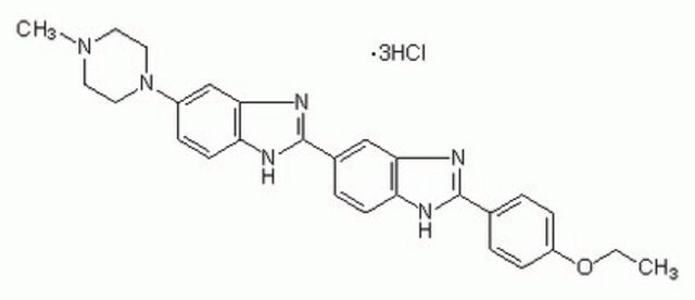
2′-(4-Ethoxyphenyl)-5-(4-methyl-1-piperazinyl)-2,5′-bi-1H-benzimidazole trihydrochloride, HOE 33342, Hoechst 33342, bisBenzimide

Sign Into View Organizational & Contract Pricing


About This Item

Empirical Formula (Hill Notation):
C27H28N6O · 3HCl · xH2O
CAS Number:
Molecular Weight:
561.93 (anhydrous basis)
Beilstein:
1234011
MDL number:
UNSPSC Code:
12171500
PubChem Substance ID:
NACRES:
NA.47

product name

bisBenzimide H 33342 trihydrochloride, ≥98% (HPLC and TLC)

Quality Level

Assay

≥98% (HPLC and TLC)

form

powder

technique(s)

titration: suitable

color

yellow

pH

1.7 (20 °C)

solubility

H2O: 20 mg/mL
phosphate buffer: precipitates

suitability

suitable for fluorescence

application(s)

diagnostic assay manufacturing
hematology
histology

storage temp.

−20°C

SMILES string

Cl[H].Cl[H].Cl[H].CCOc1ccc(cc1)C2=NCc3cc(ccc3N2)C4=NCc5cc(ccc5N4)N6CCN(C)CC6

InChI

1S/C29H32N6O.3ClH/c1-3-36-25-8-4-20(5-9-25)28-30-18-22-16-21(6-10-26(22)32-28)29-31-19-23-17-24(7-11-27(23)33-29)35-14-12-34(2)13-15-35;;;/h4-11,16-17H,3,12-15,18-19H2,1-2H3,(H,30,32)(H,31,33);3*1H

InChI key

FYEVKHPLBHLWHK-UHFFFAOYSA-N

Looking for similar products? Visit Product Comparison Guide

Application

Bisbenzimide Hoechst 33342 is a specific stain for AT-rich regions of double-stranded DNA and has been shown to displace several known DNA intercalators. This fluorescent dye has been used in sorting living cells based on DNA content, used in flow cytometry for the determination of DNA content, and for the visualization of chromatin distribution in living cells. It has been used to detect BrdU incorporation into cells and in studying the initial stages apoptosis and cellcycle distribution., Chromosomes that are dividing or replicating will not stain with this dye.
Useful for staining DNA, chromosomes and nuclei. May be used for fluorescence microscopy or flow cytometry.
Excitation max. = 346 nm
Emission max. = 460 nm

Biochem/physiol Actions

Membrane-permeable, fluorescent DNA stains with low cytotoxicity that intercalate in A-T regions of DNA.

Physical properties

Fluorescent properties of bisBenzimide H 33342:
Free dye: Excitation maximum = 340 nm, Emission maximum = 510 nm (5 mM HEPES, 10 mM NaCl, pH 7.0) DNA complex: Excitation maximum = 355 nm, Emission maximum = 465 nm (5 mM HEPES, 10 mM NaCl, pH 7.0)

Preparation Note

This product is soluble in water (50 mg/ml), yielding a clear solution. The pH of a 2% solution is 1.9. It has been observed that this material will precipitate from phosphate buffer solutions.
Aqueous solutions are stable for 1 month if kept in the dark at 2-8 °C.

related product

Product No.
Description
Pricing

Storage Class Code

11 - Combustible Solids

WGK

WGK 3

Flash Point(F)

Not applicable

Flash Point(C)

Not applicable


Choose from one of the most recent versions:

Certificates of Analysis (COA)

Lot/Batch Number

Don't see the Right Version?

If you require a particular version, you can look up a specific certificate by the Lot or Batch number.

Already Own This Product?

Find documentation for the products that you have recently purchased in the Document Library.

Visit the Document Library

Customers Also Viewed

Slide 1 of 1

1 of 1

The transforming activity of Wnt effectors correlates with their ability to induce the accumulation of mammary progenitor cells.
Liu BY, et al.
Proceedings of the National Academy of Sciences of the USA, 101, 4158-4163 (2004)
F Belloc et al.
Cytometry, 17(1), 59-65 (1994-09-01)
A flow cytometric method to detect apoptotic cells is described. This method is based on the detection of differences in chromatin condensation with Hoechst 33342 as a probe and the detection of dead cells with propidium iodide as a probe
M G Ormerod et al.
Cytometry, 14(6), 595-602 (1993-01-01)
We have recently developed a method for the separation and quantification of viable apoptotic cells without the need for permeabilisation or fixation of the cells. The method is based on the observation that apoptotic rat thymocytes fluoresce more brightly than
M Gregoire et al.
Experimental cell research, 152(1), 38-46 (1984-05-01)
Chromatin distribution was visualized in living cells with the selective DNA fluorochrome Hoechst 33342. This dye was shown to be non-toxic on the rat kangaroo PTO cell line by measuring the labelled cell growth rate. The aim of this work
D J Arndt-Jovin et al.
The journal of histochemistry and cytochemistry : official journal of the Histochemistry Society, 25(7), 585-589 (1977-07-01)
The methods for measuring the deoxyribonucleic acid content of individual mammalian cells and sorting them on the basis of this parameter have until now required fixation or other treatment which renders the cells nonviable. Using a class of bis-benzimidazole dyes

Articles

Regulation of the cell cycle involves processes crucial to the survival of a cell, including the detection and repair of genetic damage as well as the prevention of uncontrolled cell division associated with cancer. The cell cycle is a four-stage process in which the cell 1) increases in size (G1-stage), 2) copies its DNA (synthesis, S-stage), 3) prepares to divide (G2-stage), and 4) divides (mitosis, M-stage). Due to their anionic nature, nucleoside triphosphates (NTPs), the building blocks of both RNA and DNA, do not permeate cell membranes.

We presents an article on ABC Transporters and Cancer Drug Resistance

Our team of scientists has experience in all areas of research including Life Science, Material Science, Chemical Synthesis, Chromatography, Analytical and many others.

Contact Technical Service