High-Throughput Drug Screening using TrueGel3D® HTS Hydrogel Plates
Nina Berges, PhD., Lab Head Discovery Pharmacology
Merck Healthcare KGaA, Darmstadt, Germany
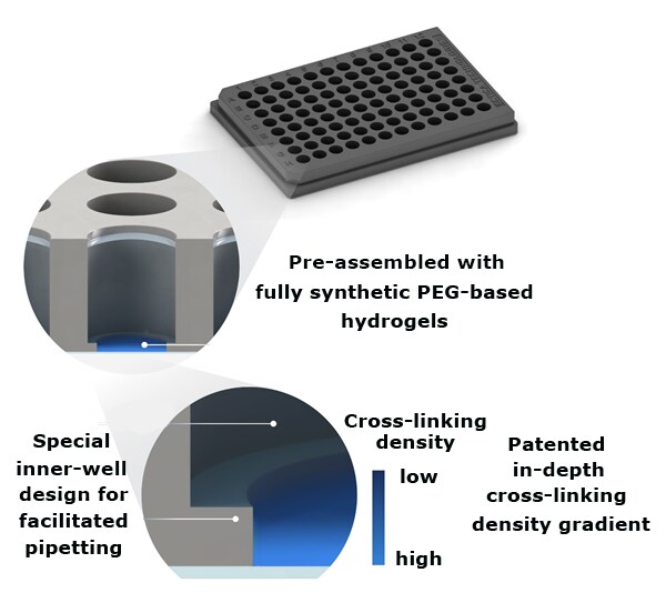
The TrueGel3D® HTS Hydrogel Plate is a ready-to-use solution to easily establish 3D cell cultures using fully synthetic hydrogels in a simple and automation-compatible manner. The 96-well polystyrene glass-bottom plates contain pre-casted synthetic functionalized PEG based hydrogels. These innovative hydrogels contain gradually increasing crosslinking densities throughout the well. Users can proceed directly with seeding cells without any hydrogel preparation or encapsulation steps. After cell seeding, cells will gradually infiltrate the hydrogel and establish a 3D cell culture environment within days.

Figure 1.TrueGel3D® HTS Hydrogel Plate technology overview. Synthetic functionalized PEG hydrogels are precast into glass bottom black polystyrene 96-well imaging plates.
Here, we utilize the TrueGel3D® HTS Hydrogel Plate to culture tumor spheroids in 3D and then screen for potential chemotherapeutic drug candidates. Using confocal imaging and a “cell burst assay” we identify compound A with high tumor cell toxicities with dose dependent effects. These data demonstrate the ability to rapidly screen large number of potential drug candidates in 3D cultures using our unique ready-to-use multiwell 3D TrueGel3D® HTS hydrogel well-plate.
Compound Cytotoxicity Screening Assay Using 3D Cell Cultures

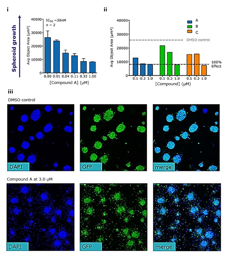
Figure 2. Spheroid growth assay detects compound cytotoxic effects of various drug compounds.EBC1-GFP spheroid cells (5k/well) grown in TrueGel3D® HTS plates treated with compounds A, B, and C show dose dependent effects with decreased spheroid size (i, ii) and increased cell death (iii) at increasing concentrations. IC50 were calculated using a VEGF-A ELISA assay (IC50 compound A: 40 nM; IC50 compound B: 550 nM; IC50 compound C; 220 nM).
Dose-dependent Cytotoxic Response Using 3D Cell Cultures

Figure 3. Dose response of compound A. EBC1-GFP spheroid size was monitored over a 3-day period after treatment with various concentrations of compound A (0 – 3 µM). Spheroids display smaller cell sizes with higher cell toxicities at higher compound A concentrations in a dose dependent manner.
3D Tumor Spheroid Killing Assay Using TrueGel3D® HTS Hydrogel Plates

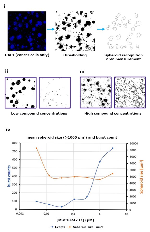
Figure 4. Spheroid killing burst assay. Spheroid killing/bursting was measured using a visual method by counting small objects (30-200 μm2 size filter) surrounding large spheroids (i). The larger number of small objects corresponds to larger cell death and toxcities. Higher concentrations of compound A (MSC1824737) leads to smaller spheroid sizes with higher cell burst counts indicating cell death (ii, iii, iv).
Pour continuer à lire, veuillez vous connecter à votre compte ou en créer un.
Vous n'avez pas de compte ?