Skip to Content
Merck
All Photos(1)

Documents

A8592

Sigma-Aldrich

Monoclonal ANTI-FLAG® M2-Peroxidase (HRP) antibody produced in mouse

clone M2, purified immunoglobulin, buffered aqueous glycerol solution

Synonym(s):

Monoclonal ANTI-FLAG® M2 antibody produced in mouse, Anti-ddddk, Anti-dykddddk

Sign Into View Organizational & Contract Pricing


About This Item

UNSPSC Code:
41106514
NACRES:
NA.32

biological source

mouse

conjugate

peroxidase conjugate

antibody form

purified immunoglobulin

antibody product type

primary antibodies

clone

M2, monoclonal

form

buffered aqueous glycerol solution

species reactivity

all

concentration

~1 mg/mL

technique(s)

indirect ELISA: 1:20,000

isotype

IgG1

immunogen sequence

DYKDDDDK

shipped in

wet ice

storage temp.

−20°C

Gene Information

human ... GPX1(2876)

Looking for similar products? Visit Product Comparison Guide

General description

The Monoclonal ANTI-FLAG M2-Peroxidase is a mouse IgG antibody covalently conjugated to horseradish peroxidase ( HRP). The antibody binds to FLAG fusion proteins and recognizes the FLAG epitope at N-terminal, Met-N-terminal, C-terminal, and internal FLAG peptides.

Application

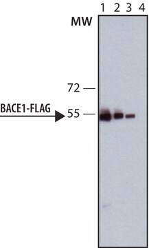
For simple, one-step detection by immunocytochemistry, ELISA, or Western blotting. Especially useful in detection of FLAG fusion proteins expressed in murine host, where secondary anti-mouse antibodies may cause cross-reactivity.
Suggested dilution for immunocytochemistry and western blotting 1:100 to 1:1000
Suggested dilution for ELISA 1:20,000

Learn more product details in our FLAG® application portal.

Physical form

Solution in phosphate buffered saline containing 50% glycerol plus preservative and stabilizer

Preparation Note

Dilute ANTI-FLAG M2-Peroxidase solution in Tris Buffered Saline (TBS): 0.05 M Tris, pH 7.4, with 0.15 M NaCl.

Legal Information

ANTI-FLAG is a registered trademark of Merck KGaA, Darmstadt, Germany
FLAG is a registered trademark of Merck KGaA, Darmstadt, Germany

Not finding the right product?  

Try our Product Selector Tool.

Signal Word

Danger

Hazard Statements

Hazard Classifications

Aquatic Acute 1 - Aquatic Chronic 1 - Eye Dam. 1 - Skin Corr. 1C - Skin Sens. 1

Storage Class Code

8A - Combustible corrosive hazardous materials

WGK

WGK 3

Flash Point(F)

Not applicable

Flash Point(C)

Not applicable


Certificates of Analysis (COA)

Search for Certificates of Analysis (COA) by entering the products Lot/Batch Number. Lot and Batch Numbers can be found on a product’s label following the words ‘Lot’ or ‘Batch’.

Already Own This Product?

Find documentation for the products that you have recently purchased in the Document Library.

Visit the Document Library

Mickaël Lelek et al.
Nature communications, 6, 6483-6483 (2015-03-07)
The molecular mechanisms that allow HIV to integrate into particular sites of the host genome are poorly understood. Here we tested if the nuclear pore complex (NPC) facilitates the targeting of HIV integration by acting on chromatin topology. We show
Dong Meng et al.
The Journal of biological chemistry, 284(17), 11425-11435 (2009-01-21)
betaArrestin is a multifunctional signal scaffold protein. Using SPOT immobilized peptide arrays, coupled with scanning alanine substitution and mutagenesis, we show that the MAPK kinase, MEK1, interacts directly with betaarrestin1. Asp(26) and Asp(29) in the N-terminal domain of betaarrestin1 are
Mai Nakashima et al.
The Journal of biological chemistry, 288(46), 33171-33180 (2013-10-08)
Continued proliferation of human cells requires maintenance of telomere length, usually accomplished by telomerase. Telomerase is recruited to chromosome ends by interaction with a patch of amino acids (the TEL patch, for TPP1 glutamate (E) and leucine (L)-rich patch) on
Gaetan Pascreau et al.
The Journal of biological chemistry, 284(9), 5497-5505 (2009-01-06)
p53 is an important tumor suppressor regulating the cell cycle at multiple stages in higher vertebrates. The p53 gene is frequently deleted or mutated in human cancers, resulting in loss of p53 activity. This leads to centrosome amplification, aneuploidy, and
Linda Brunotte et al.
Journal of virology, 85(1), 324-333 (2010-10-29)
The 200-kDa L protein of arenaviruses plays a central role in viral genome replication and transcription. This study aimed at providing evidence for the domain structure of L protein by combining bioinformatics with a stepwise mutagenesis approach using the Lassa

Articles

Structural modifications of proteins are essential to living cells. When aberrantly regulated they are often the basis of disease. Glycans are responsible for much of the structural variation in biologic systems, and their representation on cell surfaces is commonly called the “glycome.”

Our team of scientists has experience in all areas of research including Life Science, Material Science, Chemical Synthesis, Chromatography, Analytical and many others.

Contact Technical Service