Skip to Content
Merck
All Photos(1)

Key Documents

107R-1

Sigma-Aldrich

CD7 (EP132) Rabbit Monoclonal Primary Antibody

Sign Into View Organizational & Contract Pricing


About This Item

UNSPSC Code:
12352203
NACRES:
NA.41

biological source

rabbit

Quality Level

100
500

conjugate

unconjugated

antibody form

culture supernatant

antibody product type

primary antibodies

clone

EP132, monoclonal

description

(For In Vitro Diagnostic Use in Select Regions (See Chart))

form

buffered aqueous solution

species reactivity

human

packaging

bottle of 1.0 mL predilute (107R-17)
bottle of 7.0 mL predilute (107R-18)
vial of 0.1 mL concentrate (107R-14)
vial of 0.5 mL concentrate (107R-15)
vial of 1.0 mL concentrate (107R-16)

manufacturer/tradename

Cell Marque

technique(s)

immunohistochemistry (formalin-fixed, paraffin-embedded sections): 1:100-1:500

isotype

IgG

shipped in

wet ice

storage temp.

2-8°C

Gene Information

human ... CD7(924)

General description

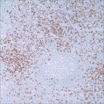
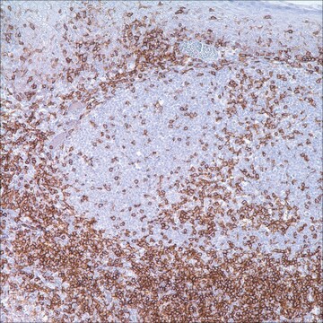
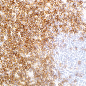
CD7 antigen is a cell surface glycoprotein of 40 kD expressed on the surface of immature and mature T-cells as well as natural Killer (NK) cells. It is a member of the immunoglobulin gene superfamily and is the first T-cell lineage associated antigen to appear in T-cell ontogeny, being expressed in T-cell precursors (preceding CD2 expression), and in myeloid precursors, in fetal liver and bone marrow, and persisting in circulating mature T-cells. While its precise function is not known, it is suggested that the molecule functions as an Fc receptor for IgM. CD7 is the most consistently expressed T-cell antigen in lymphoblastic lymphomas/leukemias, and is therefore anti-CD7 is a useful marker in the identification of such neoplastic proliferations. In mature post-thymic T-cell neoplasms, CD7 is the most common pan-T-antigen to be aberrantly expressed, which is a useful pointer to a neoplastic T-cell process. 2,3 CD7 has been shown to be immunoexpressed in 85% of mature peripheral T-cells, the majority of post-thymic T-cells, NK cells, T-cell lymphoblastic leukemia/lymphoma, acute myeloid leukemia, and chronic myelogenous leukemia, CD7 is conspicuously absent in adult T-cell leukemia/lymphoma and is not expressed in mycosis fungoides. 1-3

Quality


IVD

IVD

IVD

RUO

Linkage

CD7 Positive Control Slides, Product No. 107S, are available for immunohistochemistry (formalin-fixed, paraffin-embedded sections).

Physical form

Solution in Tris Buffer, pH 7.3-7.7, with 1% BSA and <0.1% Sodium Azide.

Preparation Note

Download the IFU specific to your product lot and formatNote: This requires a keycode which can be found on your packaging or product label.

Other Notes

For Technical Service please contact: 800-665-7284 or email: service@cellmarque.com

Legal Information

Cell Marque is a trademark of Merck KGaA, Darmstadt, Germany

Not finding the right product?  

Try our Product Selector Tool.


Choose from one of the most recent versions:

Certificates of Analysis (COA)

Lot/Batch Number

Don't see the Right Version?

If you require a particular version, you can look up a specific certificate by the Lot or Batch number.

Already Own This Product?

Find documentation for the products that you have recently purchased in the Document Library.

Visit the Document Library

Philip Went et al.
Journal of clinical oncology : official journal of the American Society of Clinical Oncology, 24(16), 2472-2479 (2006-04-26)
Although peripheral T-cell lymphoma, unspecified (PTCL/U), is the most common T-cell tumor in Western countries, no study to date has been based on the application of a wide panel of markers to a large series of patients and assessed the
Emmilia Hodak et al.
Journal of the American Academy of Dermatology, 55(2), 276-284 (2006-07-18)
Mycosis fungoides (MF) is an epidermotropic cutaneous T-cell lymphoma in which the tumor cells express a mature T-helper memory phenotype, ie, CD3(+), CD4(+), CD8(-), CD45RO(+), with a T-cell receptor (TCR) of the alpha/beta heterodimer. A minority of patients have an
Eric C Vonderheid
Journal of cutaneous pathology, 33 Suppl 1, 27-42 (2006-01-18)
Erythrodermic cutaneous T-cell lymphoma (E-CTCL) is the cause of less than 5% of all cases of generalized erythroderma. A methodical evaluation of skin, blood, and lymph node samples using standard histology, immunohistochemistry (IHC), flow cytometry (FC), and molecular analysis for

Our team of scientists has experience in all areas of research including Life Science, Material Science, Chemical Synthesis, Chromatography, Analytical and many others.

Contact Technical Service