Skip to Content
Merck
All Photos(2)

Key Documents

ABE1957

Sigma-Aldrich

Anti-CENP-C Antibody

serum, from rabbit

Synonym(s):

Centromere protein C, CENP-C, CENP-C 1, Centromere autoantigen C, Centromere protein C 1, Interphase centromere complex protein 7

Sign Into View Organizational & Contract Pricing


About This Item

UNSPSC Code:
12352203
eCl@ss:
32160702

biological source

rabbit

Quality Level

antibody form

serum

antibody product type

primary antibodies

clone

polyclonal

species reactivity

human

technique(s)

immunocytochemistry: suitable
western blot: suitable

NCBI accession no.

UniProt accession no.

shipped in

ambient

target post-translational modification

unmodified

Gene Information

human ... CENPC(1060)

General description

Centromere protein C (UniProt Q03188; also known as CENP-C, CENP-C 1, Centromere autoantigen C, Centromere protein C 1, Interphase centromere complex protein 7) is encoded by the CENPC (also known as CENPC1, ICEN7) gene (Gene ID 1060) in human. Kinetochores assemble on centromeres to bind spindle microtubules and mediate chromosome segregation during cell division. CENP-C is a subunit of constitutive centromere-associated network (CCAN) that acts as the centromere-kinetochore interface. CCAN is a large complex composed of at least 16 different centromeric proteins (CENPs). CENP-C plays an important role in kinetochore assembly downstream of CENP-A. The PEST domain in the N-terminal half of CENP-C interacts directly with the CCAN subcomplex CENP-HIKM (CENP-H, CENP-K, CENP-I, and CENP-M) and CENP-C depletion is shown to cause mislocalization of CENP-T and the CENP-HIKM complex subunits in HeLa cells.

Specificity

This rabbit polyclonal antiserum targets the N-terminal region of human CENP-C present in both spliced isoforms reported by UniProt (Q03188). Specificity was demonstrated by a reducion of target band detection (Western blotting) and kinetochores staining (chromsome spreads fluoresence staining) following cellular CENP-C shRNA induction (Falk, S.J., et al. (2015). Science. 348(6235):699-703).

Immunogen

Epitope: N-terminus
GST-tagged recombinant human CENP-C N-terminal fragment.

Application

Anti-CENP-C Antibody, Cat. No. ABE1957, is a highly specific rabbit polyclonal antibody that targets CENP-C and has been tested in Immunocytochemistry and Western Blotting.
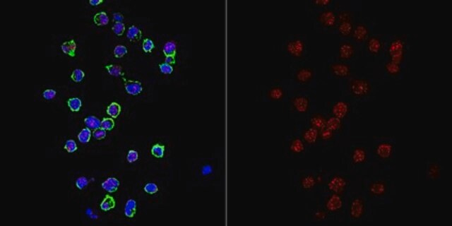
Immunocytochemistry Analysis: A 1:10,000 dilution from a representative lot immunostained kinetochores in chromosome spreads prepared from interphase DLD-1 human colorectal adenocarcinoma cells hypotonically swollen and fixed with 4% formaldehyde.

Immunocytochemistry Analysis: A representative lot was affinity purified and detected a time-dependent loss of kinetochores CENP-C immunoreactivity in 4% formaldehyde-fixed, 0.5% Triton X-100-permeabilized HeLa cells following CENP-C shRNA induction (Falk, S.J., et al. (2015). Science. 348(6235):699-703).

Immunocytochemistry Analysis: A representative lot was affinity purified and immunostained kinetochores by indirect fluorescence staining of chromosome spreads prepared from patient-derived, PD-NC4 chromosome variant harboring fibroblasts hypotonically swollen and fixed with 4% formaldehyde (Bassett, E.A., et al. (2010). J. Cell Biol. 190(2):177-185).

Western Blotting Analysis: A representative lot was affinity purified and detected a time-dependent CENP-C level in HeLa cells following CENP-C shRNA induction (Falk, S.J., et al. (2015). Science. 348(6235):699-703).
Research Category
Epigenetics & Nuclear Function

Quality

Evaluated by Western Blotting in DLD-1 cell lysate.

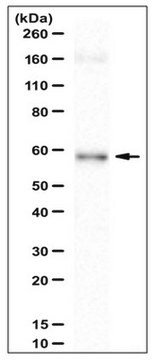
Western Blotting Analysis: A 1:5,000 dilution of this antiserum detected CENP-C in 50,000 cell equivalent of DLD-1 human colorectal adenocarcinoma cell lysate.

Target description

~140 kDa observed. 106.8 kDa (isoform 1) and 61.54 kDa (isoform 2) calculated. The larger-than-calculated target band size is consistent with that reported in the literature (Earnshaw, W.C., et al. (1989). Chromosoma. 98(1):1-12). Uncharacterized bands may be observed in some lysate(s).

Physical form

Rabbit polyclonal antibody serum with 0.05% sodium azide.
Unpurified.

Storage and Stability

Stable for 1 year at -20°C from date of receipt.
Handling Recommendations: Upon receipt and prior to removing the cap, centrifuge the vial and gently mix the solution. Aliquot into microcentrifuge tubes and store at -20°C. Avoid repeated freeze/thaw cycles, which may damage IgG and affect product performance.

Other Notes

Concentration: Please refer to lot specific datasheet.

Disclaimer

Unless otherwise stated in our catalog or other company documentation accompanying the product(s), our products are intended for research use only and are not to be used for any other purpose, which includes but is not limited to, unauthorized commercial uses, in vitro diagnostic uses, ex vivo or in vivo therapeutic uses or any type of consumption or application to humans or animals.

Not finding the right product?  

Try our Product Selector Tool.

Storage Class Code

12 - Non Combustible Liquids

WGK

WGK 1

Flash Point(F)

Not applicable

Flash Point(C)

Not applicable


Certificates of Analysis (COA)

Search for Certificates of Analysis (COA) by entering the products Lot/Batch Number. Lot and Batch Numbers can be found on a product’s label following the words ‘Lot’ or ‘Batch’.

Already Own This Product?

Find documentation for the products that you have recently purchased in the Document Library.

Visit the Document Library

Our team of scientists has experience in all areas of research including Life Science, Material Science, Chemical Synthesis, Chromatography, Analytical and many others.

Contact Technical Service