Skip to Content
Merck
All Photos(4)

Key Documents

AMAB90873

Sigma-Aldrich

Monoclonal Anti-CD68 antibody produced in mouse

Prestige Antibodies® Powered by Atlas Antibodies, clone CL1338, purified immunoglobulin, buffered aqueous glycerol solution

Synonym(s):

Anti-DKFZp686M18236, Anti-GP110, Anti-LAMP4, Anti-SCARD1, Anti-macrosialin

Sign Into View Organizational & Contract Pricing


About This Item

UNSPSC Code:
12352203
NACRES:
NA.41

Pricing and availability is not currently available.

biological source

mouse

Quality Level

conjugate

unconjugated

antibody form

purified immunoglobulin

antibody product type

primary antibodies

clone

CL1338, monoclonal

product line

Prestige Antibodies® Powered by Atlas Antibodies

form

buffered aqueous glycerol solution

species reactivity

human

technique(s)

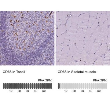
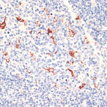
immunohistochemistry: 1:1000- 1:2500

Compare Similar Items

View Full Comparison

Show Differences

1 of 4

This Item
1392002PHR1611BP762
manufacturer/tradename

BP

manufacturer/tradename

USP

manufacturer/tradename

-

manufacturer/tradename

BP

format

neat

format

neat

format

neat

format

mixture

application(s)

pharmaceutical
pharmaceutical small molecule

application(s)

pharmaceutical (small molecule)

application(s)

pharmaceutical (small molecule)

application(s)

pharmaceutical
pharmaceutical small molecule

form

solid

form

-

form

-

form

solid

shelf life

limited shelf life, expiry date on the label

shelf life

-

shelf life

-

shelf life

limited shelf life, expiry date on the label

Immunogen

CD68 molecule

Application

All Prestige Antibodies Powered by Atlas Antibodies are developed and validated by the Human Protein Atlas (HPA) project and as a result, are supported by the most extensive characterization in the industry.

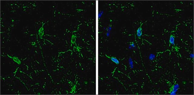
The Human Protein Atlas project can be subdivided into three efforts: Human Tissue Atlas, Cancer Atlas, and Human Cell Atlas. The antibodies that have been generated in support of the Tissue and Cancer Atlas projects have been tested by immunohistochemistry against hundreds of normal and disease tissues and through the recent efforts of the Human Cell Atlas project, many have been characterized by immunofluorescence to map the human proteome not only at the tissue level but now at the subcellular level. These images and the collection of this vast data set can be viewed on the Human Protein Atlas (HPA) site by clicking on the Image Gallery link. We also provide Prestige Antibodies® protocols and other useful information.
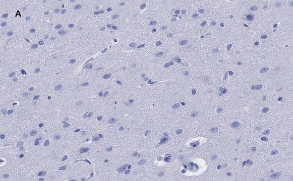
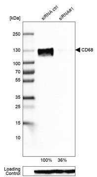
Monoclonal Anti-CD68 Prestige Antibodies® Powered by Atlas Antibodies is developed and validated by the Human Protein Atlas (HPA) project . Each antibody is tested by immunohistochemistry against hundreds of normal and disease tissues. These images can be viewed on the Human Protein Atlas (HPA) site by clicking on the Image Gallery link. The antibodies are also tested using protein array and western blotting. To view these protocols and other useful information about Prestige Antibodies and the HPA, visit sigma.com/ Prestige.

Features and Benefits

Prestige Antibodies® are highly characterized and extensively validated antibodies with the added benefit of all available characterization data for each target being accessible via the Human Protein Atlas portal linked just below the product name at the top of this page. The uniqueness and low cross-reactivity of the Prestige Antibodies® to other proteins are due to a thorough selection of antigen regions, affinity purification, and stringent selection. Prestige antigen controls are available for every corresponding Prestige Antibody and can be found in the linkage section.

Every Prestige Antibody is tested in the following ways:
  • IHC tissue array of 44 normal human tissues and 20 of the most common cancer type tissues.
  • Protein array of 364 human recombinant protein fragments.

Linkage

Corresponding Antigen APREST85088

Physical form

Phospate buffered saline, pH 7.2, containing 40% glycerol and 0.02% sodium azide

Legal Information

Prestige Antibodies is a registered trademark of Merck KGaA, Darmstadt, Germany

Disclaimer

Unless otherwise stated in our catalog or other company documentation accompanying the product(s), our products are intended for research use only and are not to be used for any other purpose, which includes but is not limited to, unauthorized commercial uses, in vitro diagnostic uses, ex vivo or in vivo therapeutic uses or any type of consumption or application to humans or animals.

Not finding the right product?  

Try our Product Selector Tool.

Storage Class Code

10 - Combustible liquids

WGK

WGK 1

Flash Point(F)

Not applicable

Flash Point(C)

Not applicable


  • Choose from one of the most recent versions:

    Certificates of Analysis (COA)

    Lot/Batch Number

    Sorry, we don't have COAs for this product available online at this time.

    If you need assistance, please contact Customer Support.

    Already Own This Product?

    Find documentation for the products that you have recently purchased in the Document Library.

    Visit the Document Library

    William B Laskin et al.
    The American journal of surgical pathology, 38(1), 1-12 (2013-10-15)
    In this study, we examine the clinicopathologic features of 104 cases of myxoinflammatory fibroblastic sarcoma (MIFS), a low-grade, inflammatory fibromyxoid tumor with a predilection to distal extremity soft tissue, and attempt to identify factors predictive of aggressive behavior. The study
    Shogo Kumagai et al.
    Lung cancer (Amsterdam, Netherlands), 85(3), 457-464 (2014-07-22)
    Increasing evidence suggests that an elevated peripheral monocyte count at presentation predicts a poor prognosis in various types of malignancy, including malignant lymphoma. In lung adenocarcinoma, tumor-associated macrophages (TAMs) were reported to be associated with a poor prognosis. However, it
    Jing Wang et al.
    Biochimica et biophysica acta, 1842(11), 2174-2183 (2014-08-06)
    Cathepsin G (CatG), a serine protease present in mast cells and neutrophils, can produce angiotensin-II (Ang-II) and degrade elastin. Here we demonstrate increased CatG expression in smooth muscle cells (SMCs), endothelial cells (ECs), macrophages, and T cells from human atherosclerotic
    Manabu Shigeoka et al.
    Cancer science, 104(8), 1112-1119 (2013-05-08)
    Tumor associated macrophages (TAMs) are the most abundant cancer stromal cells educated by tumor microenvironment to acquire trophic functions facilitating angiogenesis, matrix breakdown and cancer cell motility. Tumor associated macrophages have anti-inflammatory properties or "alternatively" activated (M2) phenotype expressing CD204
    Jenny Sandström von Tobel et al.
    Neurotoxicology, 44, 61-70 (2014-05-06)
    Neurotoxic effects of the environmentally abundant mycotoxin Ochratoxin A (OTA) were studied in histotypic 3D rat brain cell cultures, comprising all brain cell types. Cultures were exposed to nanomolar OTA concentrations and samples were collected 48h after a single exposure

    Our team of scientists has experience in all areas of research including Life Science, Material Science, Chemical Synthesis, Chromatography, Analytical and many others.

    Contact Technical Service