AB5535
Anti-SOX9 Antibody
CHEMICON®, rabbit polyclonal
Synonym(s):
Anti-CMD1, Anti-CMPD1, Anti-SRA1, Anti-SRXX2, Anti-SRXY10
About This Item
Recommended Products
Product Name
Anti-Sox9 Antibody, Chemicon®, from rabbit
biological source
rabbit
Quality Level
antibody form
affinity isolated antibody
antibody product type
primary antibodies
clone
polyclonal
purified by
affinity chromatography
species reactivity
chicken, human, rat, mouse
species reactivity (predicted by homology)
bovine (based on 100% sequence homology), sheep (based on 100% sequence homology), feline (based on 100% sequence homology), equine (based on 100% sequence homology)
packaging
antibody small pack of 25 μg
manufacturer/tradename
Chemicon®
technique(s)
ChIP: suitable (ChIP-seq)
immunocytochemistry: suitable
immunofluorescence: suitable
immunohistochemistry: suitable
western blot: suitable
NCBI accession no.
UniProt accession no.
shipped in
ambient
storage temp.
2-8°C
target post-translational modification
unmodified
Gene Information
human ... SOX9(6662) , SRA1(10011)
General description
Specificity
Immunogen
Application
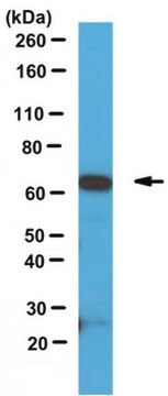
Western Blotting Analysis: An 1:500 dilution from a representative lot detected Sox9 in human PC3 prostate cancer cells and HepG2 hepatocytes.
Chromatin Immunoprecipitation (ChIP) Analysis: A representative lot detected Sox9 occupancy at target chromatin sites by ChIP using chromatin preparations from P1 post-natal mouse rib chondrocytes (Ohba, S., et al. (2015). Cell Rep. 12(2):229-243).
Chromatin Immunoprecipitation (ChIP) Analysis: A representative lot detected Sox9 occupancy at the Bmi promoter in Z/sox9tg but not in wild-type control mouse embryonic fibroblasts/MEFs (Matheu, A., et al. (2012). Cancer Res. 72(5):1301-1315).
ChIP-sequencing (ChIP-seq) Analysis: A representative lot detected Sox9-targeted chromatin sites by a genome-wide ChIP-seq analysis using chromatin preparations from P1 post-natal mouse rib chondrocytes (Ohba, S., et al. (2015). Cell Rep. 12(2):229-243).
Immunofluorescence Analysis: A representative lot detected the accumulation of Sox9-positive oval cells by fluorescent immunohistochemistry staining of paraffin-embedded liver sections from transgenic mice treated with diethylnitrosamine to induce conditional liver HNF4a knockout (Saha, S.K., et al. (2014). Nature. 513(7516):110-114).
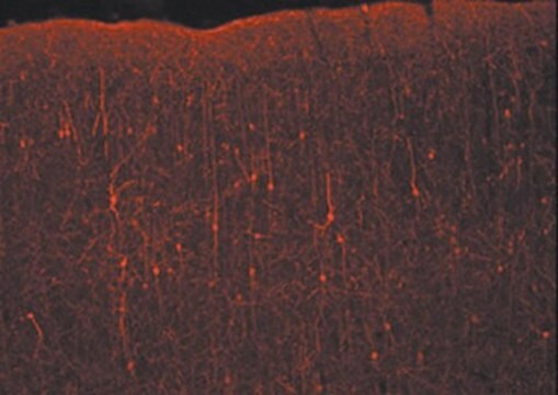
Immunofluorescence Analysis: Representative lots detected Sox9 immunoreactivity in paraffin-embedded mouse embryo sections by fluorescent immunohistochemistry (Carrasco, M., et al. (2012). J. Clin. Invest. 122(10):3504-3515; Sylva, M., et al. (2011). PLoS One. 6(8):e22616).
Immunofluorescence Analysis: Representative lots immunostained Müller glial cells in frozen mouse and chicken retina sections by fluorescent immunohistochemistry staining of (Muranishi, Y., and Furukawa, T. (2012). J. Biomed. Biotechnol. 2012:973140; Fischer, A.J., et al. (2011). Neuroscience. 178:250-260).
Immunocytochemistry Analysis: A representative lot detected the stem cell marker Sox9 by fluorescent immunocytochemistry staining of paraformaldehyde-fixed E-Cad/Lgr6+ human lung stem cells (HLSCs) clonally derived and passaged in culture (Oeztuerk-Winder, F., et al. (2012). EMBO J. 31(16):3431-3441).
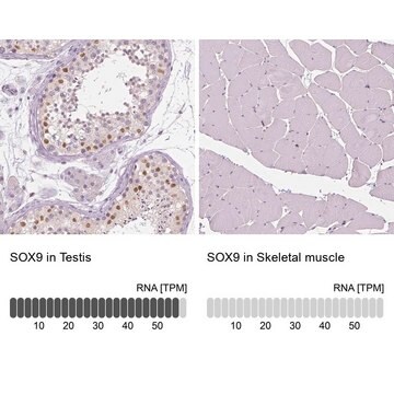
Immunohistochemistry Analysis: A representative lot immunostained the supporting cells (Sertoli) of the seminiferous tubules by immunohistochemistry staining of paraffin-embedded mouse testis sections (O′Shaughnessy, P.J., et al. (2012). PLoS One. 7(4):e35136).
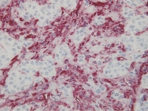
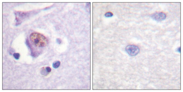
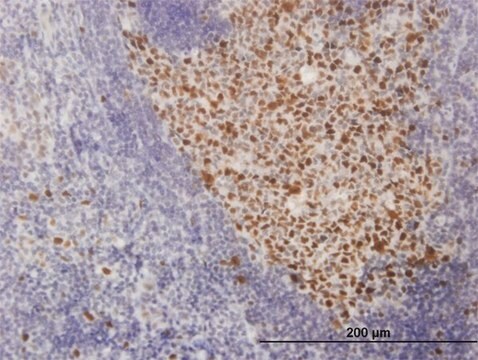
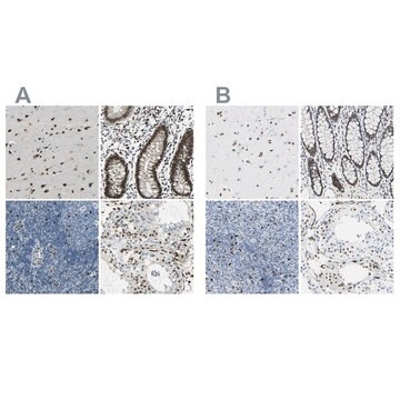
Immunohistochemistry Analysis: A representative lot detected Sox9 immunoreactivity in various formalin-fixed, paraffin-embedded human tumor tissue sections (Matheu, A., et al. (2012). Cancer Res. 72(5):1301-1315).
Western Blotting Analysis: A representative lot detected the stem cell marker Sox9 in E-Cad/Lgr6+ human lung stem cells (HLSCs) clonally derived and passaged in culture (Oeztuerk-Winder, F., et al. (2012). EMBO J. 31(16):3431-3441).
Western Blotting Analysis: A representative lot detected upregulated Sox9 expression level in human colorectal cancer cell lines, HCT116, DLD1, and SW620 (Matheu, A., et al. (2012). Cancer Res. 72(5):1301-1315).
Epigenetics & Nuclear Function
Transcription Factors
Quality
Western Blotting Analysis: An 1:2000 dilution of this antibody detected Sox9 in HepG2 cell lysate.
Target description
Linkage
Physical form
Storage and Stability
Analysis Note
Embryonic tissue, Adult chondrocytes.
Other Notes
Legal Information
Disclaimer
Not finding the right product?
Try our Product Selector Tool.
recommended
Storage Class Code
12 - Non Combustible Liquids
WGK
WGK 2
Flash Point(F)
Not applicable
Flash Point(C)
Not applicable
Certificates of Analysis (COA)
Search for Certificates of Analysis (COA) by entering the products Lot/Batch Number. Lot and Batch Numbers can be found on a product’s label following the words ‘Lot’ or ‘Batch’.
Already Own This Product?
Find documentation for the products that you have recently purchased in the Document Library.
Customers Also Viewed
Protocols
How to stain organoids? A complete step-by-step protocol for immunofluorescent (IF) and immunocytochemical (ICC) staining of organoid cultures using antibodies
Our team of scientists has experience in all areas of research including Life Science, Material Science, Chemical Synthesis, Chromatography, Analytical and many others.
Contact Technical Service