Skip to Content
Merck
All Photos(3)

Key Documents

06-427

Sigma-Aldrich

Anti-Phosphotyrosine Antibody

Upstate®, from rabbit

Sign Into View Organizational & Contract Pricing


About This Item

UNSPSC Code:
12352203
eCl@ss:
32160702
NACRES:
NA.41

biological source

rabbit

Quality Level

antibody form

affinity purified immunoglobulin

antibody product type

primary antibodies

clone

polyclonal

purified by

affinity chromatography

species reactivity

human

species reactivity (predicted by homology)

all (based on 100% sequence homology)

manufacturer/tradename

Upstate®

technique(s)

immunoprecipitation (IP): suitable
western blot: suitable

shipped in

wet ice

target post-translational modification

acetylation (test)

Gene Information

human ... PID1(55022)

General description

The phosphorylation of specific tyrosine residues has been shown to be a primary mechanism of signal transduction during normal mitogenesis, cell cycle progression and oncogenic transformation, its role in other areas such as differentiation and gap junction communication, is a matter of active and ongoing research. Antibodies that specifically recognize phosphorylated tyrosine residues have proved to be invaluable to the study of tyrosine phosphorylated proteins and the biochemical pathways in which they function.

Specificity

The immunoreactivity of the antibody is totally inhibited by the use of 100 mM phenyl phosphate, a phosphotyrosine analog. The phosphotyrosine antibody is purified by immunoaffinity chromatography using either a dual phospho-peptide gel or a BSA-phosphotyrosine gel. All of the phosphotyrosine immunoreactivity present in the antisera is immunoadsorbed whether the antibody is purified by either gel indicating that the antibody is not sequence-specific but specific for phosphotyrosine residues.

Immunogen

In order to produce broad spectrum polyclonal phosphotyrosine antibodies, rabbits were immunized with three phosphorylated immunogens: (1) phosphotyrosine covalently linked to KLH; (2) the c-Src carboxyl terminal regulatory phosphopeptide (TS-T-E-P-Q-pY-Q-P-G-E-N-L; Catalog # 12-218) covalently linked to KLH, and; (3) a phosphopeptide associated with high tyrosine kinase activity in human lymphocytes (R-R-L-I-E-D-A-E-pY-A-A-R-G; Catalog # 12-217) covalently linked to KLH. Both of the phosphopeptide haptens serve as strong substrates for tyrosine phosphatases and are part of the two colorimetric tyrosine phosphatase kits provided by Upstate, Inc. (Catalog # 17-125, 17-126).

Application

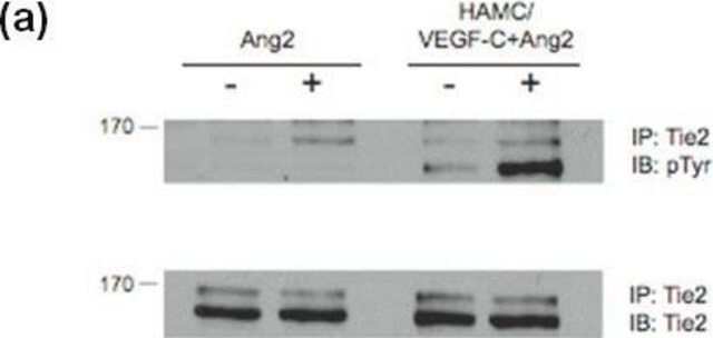
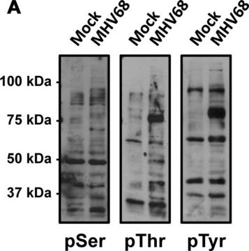
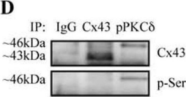
Anti-Phosphotyrosine Antibody detects level of Phosphotyrosine & has been published & validated for use in IP & WB.
Research Category
Signaling
Research Sub Category
General Post-translation Modification

Signaling Neuroscience
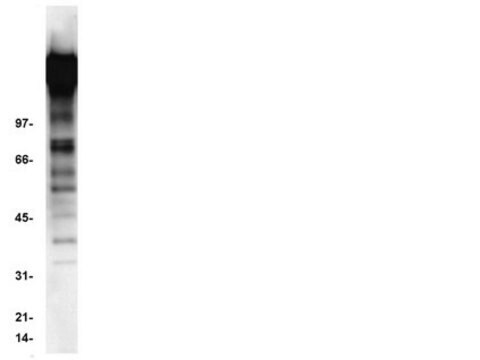
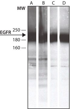
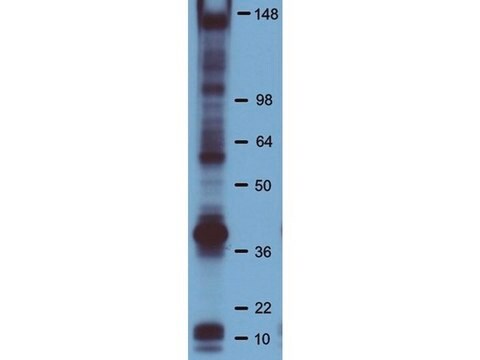
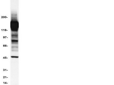
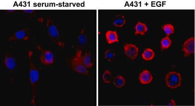
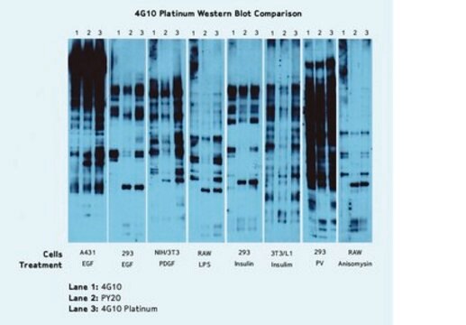
Western Blotting Analysis: A 1:500 dilution from a representative lot detected Phosphotyrosine in 10 µg of EGF treated A431 with Lambda Phosphatase tissue lysate.
Immunoprecipitation Analysis: 5 µL from a representative lot immunoprecipitated Phosphotyrosine in 0.5 mg of EGF treated A431 cell lysate

Quality

Evaluated by Western Blotting in EGF treated A431 cell lysate.

Western Blotting Analysis: A 1:500 dilution of this antibody detected Phosphotyrosine in 10 µg of EGF treated A431 cell lysate.

Target description

Varies

Physical form

Affinity purified
Purified rabbit polyclonal in buffer containing 0.1 M Tris-Glycine (pH 7.4), 150 mM NaCl with 0.05% sodium azide.

Storage and Stability

Stable for 1 year at 2-8°C from date of receipt.

Analysis Note

Control
Positive Antigen Control: Catalog #12-302, EGF-stimulated A431 cell lysate. Add 2.5µL of 2-mercaptoethanol/100µL of lysate and boil for 5 minutes to reduce the preparation. Load 20µg of reduced lysate per lane for minigels.

Other Notes

Concentration: Please refer to the Certificate of Analysis for the lot-specific concentration.

Legal Information

UPSTATE is a registered trademark of Merck KGaA, Darmstadt, Germany

Disclaimer

Unless otherwise stated in our catalog or other company documentation accompanying the product(s), our products are intended for research use only and are not to be used for any other purpose, which includes but is not limited to, unauthorized commercial uses, in vitro diagnostic uses, ex vivo or in vivo therapeutic uses or any type of consumption or application to humans or animals.

Not finding the right product?  

Try our Product Selector Tool.


Certificates of Analysis (COA)

Search for Certificates of Analysis (COA) by entering the products Lot/Batch Number. Lot and Batch Numbers can be found on a product’s label following the words ‘Lot’ or ‘Batch’.

Already Own This Product?

Find documentation for the products that you have recently purchased in the Document Library.

Visit the Document Library

Wnt5a is required for proper mammary gland development and TGF-beta-mediated inhibition of ductal growth.
Roarty, K; Serra, R
Development null
Selective activation of T cell kinase p56lck by Herpesvirus saimiri protein tip
Wiese, N., et al
The Journal of Biological Chemistry, 271, 847-852 (1996)
In vivo insulin signaling through PI3-kinase is impaired in skeletal muscle of adult rat offspring exposed to ethanol in utero.
Chen, L; Yao, XH; Nyomba, BL
Journal of Applied Physiology (1985)
Human papillomavirus type 6b virus-like particles are able to activate the Ras-MAP kinase pathway and induce cell proliferation
Payne, E., et al
Journal of virology, 75, 4150-4157 (2001)
Phosphoinositide 3-kinase, Src, and Akt modulate acute ventilation-induced vascular permeability increases in mouse lungs.
Miyahara, T; Hamanaka, K; Weber, DS; Drake, DA; Anghelescu, M; Parker, JC
American Journal of Physiology. Lung Cellular and Molecular Physiology null

Our team of scientists has experience in all areas of research including Life Science, Material Science, Chemical Synthesis, Chromatography, Analytical and many others.

Contact Technical Service