Guidelines for Bioassays on MultiScreen Filter Plates: Protein Binding and Cell Loading

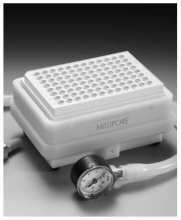
The MultiScreenHTS 96-well filter plate
Optimization Parameters for Receptor Binding, Enzyme Activity, Drug Discovery and Related Assays
The MultiScreen Assay System is well suited for a variety of separation techniques, including cell separations and solid phase bioassays. The MultiScreen system makes it possible to carry out an entire assay from sample incubation to scintillation counting in a single 96-well plate. The advantages of the system include a 96 % or more reduction in radioactive waste over traditional cell harvesting methods, improved coefficients of variation, better assay reproducibility, and faster multiple sample processing. Most assays carried out using historical methods and techniques can now be carried out on one of the different filter media available on the MultiScreen 96-well plates. To ensure accurate, reliable assays, certain technical parameters should be observed depending on the type of MultiScreen plate you select. These issues are particularly important when “miniaturizing” an assay, or when optimizing an established method with the actual 96-well plate. These guidelines will help you select the optimal filter for a specific application.
For assays where binding capacity required exceeds that of membranes, the Durapore® plates loaded with suitable chromatography media are recommended. Use of plates containing glass fiber or filter papers is not recommended when adding resins or beads. Increased dead volume, higher non-specific binding and sometimes reduced filtration capacity are caused by interactions between filter materials and media. Plates are available for both solvent-based and aqueous applications.
In addition to selecting the right membrane material for your assay, you must also make sure to select the right plate material. Table 1 for recommendations.
In some of the following tables, not all of the available MultiScreen membrane filters will have data, since some filters are inappropriate for certain techniques. The five technical parameters addressed are: 1) plate preparation, 2) protein binding, 3) protein (IgG) loading or membrane capacity, 4) cell loading, and 5) bacteria loading.
Materials and Methods for Guidelines Testing
Methods for determining experimental parameters for the different MultiScreen plates are described in the appropriate sections. The materials listed below were used in the various studies. Proteins and 125I labeled proteins were purchased from Sigma Chemical Co. (St. Louis, MO), ICN (Irvine, CA) and NEN (Boston, MA).

Solvent-resistant MultiScreenHTS vacuum manifold
MultiScreen Plate Preparation and Punching Recommendations
Prior to the addition of assay reagents, the individual wells of the MultiScreen plate may require pre-wetting for optimal assay performance. Table 3, column 3, shows the recommended pre-wet. To pre-wet the plate, add appropriate liquid, incubate for one minute, filter through using the MultiScreen or other vacuum manifold. Some membrane types can be punched into scintillation or gamma vials using the MultiScreen Multiple Punch. Table 3, column 4, shows punching capability. MultiScreen opaque plates are recommended for direct detection with microplate scintillation or luminescent counters.
Protein Binding Guidelines
Protein binding levels vary directly with the type of membrane media in the MultiScreen plate1. For high protein binding applications use IP, HA, DE, or PH plates. For high nucleic acid binding use HA, FB, FC, or DE plates. For low biomolecule binding use Durapore plates. For assays that require solvent resistance, use RL or RP Solvinert filter plates. The FL plates are recommended solely for cell culture assays. General guidelines for protein binding are reported in Table 4.
1Protein eluted in 0.5 M NaCl for total protein assay
2ND = Not determined
The MultiScreen-FC and FB glass fiber filter plates show high binding capacity for BSA and IgG, but low binding capacity for insulin. These plates are primarily intended for receptor binding assays, particularly with crude receptor preparations. The FB plates are also suitable for DNA binding.
The plates containing Durapore membranes, including GV, HV, DV and BV, all demonstrate low protein binding. These membranes have excellent nonspecific binding levels (NSB) and are recommended for column separation assays, enzyme assays, and biological sample preparation in aqueous conditions. These MultiScreen plates are used extensively for cytokines such as interleukins2,4,5 and 6. They are also ideal for filtering multiple small volume samples directly into plastic 96-well plates.
Each microporous membrane interacts distinctly with each protein. For this reason, it may be necessary to test different membrane types for each assay. The MultiScreen Assay System Applications and References Guide (RF016) recommends plates by assay type, and lists references to published applications.
Procedure
The following procedure was used to determine protein binding levels for various media:
Plates (with the exception of IP) were pre-wet with 200 µL phosphate buffered saline with 0.01 % sodium azide, vacuum drained, and then the underdrain was blotted on clean paper towels. The IP plate was first pre-wet with 20 µL of 50 % ethanol, vacuum drained, and then washed three times with 200 µL PBS and blotted on paper towels.
Using a multichannel pipette,100 µL of 125I-BSA, 125I-IgG or 125I-insulin was added into one row (8 wells) on each plate. Each 125I-labeled protein solution was a 1 mg/mL solution with approximately 105 CPM per 100 µL. Plates were incubated for two hours at room temperature, vacuum drained, washed and vacuum drained four times with 200 µL of PBS, and blotted on paper towels. The underdrain was removed and the membrane blotted on paper towels. Plates were punched using the MultiScreen punch assembly into 12 x 75 mm test tubes.
Gamma counting was carried out on each membrane using Packard’s Minaxi-γ Auto-Gamma® 5000 Series.
Protein Loading Recommendations
For optimum assay sensitivity, the loading levels of protein for each well of the MultiScreen plate should be kept at a level that maximizes effective washing. The loading guidelines noted in Table 4 below indicate general protein loading levels based on studies of TCA-precipitated IgG. The criteria for recommended maximum loading was the capacity to perform each wash at 12" Hg vacuum in less than 20 seconds.
To determine protein loading levels, plates were first pre-wet, as in Table 3. The first column of the plate was loaded with a 10 % IgG solution and serial diluted in PBS across the plates to achieve a range of dilutions. Next, 100 µL of cold 20 % trichloroacetic acid (TCA) was added to all wells on all plates. Plates were incubated at 4 °C for 30 minutes and vacuum drained. Plates were then washed three times with 10 % cold TCA.
Cell Loading Recommendations
Cell loading studies were performed on the MultiScreen plates to determine the maximum level of cells per well that can be filtered and washed efficiently. These recommendations are useful for cell-based assays involving crude receptor preparations or cell culture suspensions. The recommended guidelines for cell loading on appropriate MultiScreen media are shown in Table 5. Cell loading studies on the various MultiScreen plates were carried out with mouse hybridomas (ATCC #TIB-111). Cells were grown and resuspended to a final concentration of 5 x 10⁶ cells/150 µL. After pre-wetting the plate, the first column was loaded with 150 µL of sample.
Serial dilutions were made in 150 µL of media across the plate to achieve desired cell loads. After incubation, vacuum was applied and the plates washed three times with PBS. Criteria for maximum recommended loading was the ability to accomplish each of the three washes at 12" Hg vacuum in less than 20 seconds. Recommendations for receptor preparations are based on this data and filtration capacities for the membranes.
Seeding Density Recommendations
Seeding density is the recommended number of cells/well to load onto a plate for cell culture, e.g. for subsequent cell proliferation assays.
1Mouse hybridoma cells
2NR = Not recommended
Bacteria Loading Recommendations
For sample preparation of bacteria, a procedure that maximizes effective washing will maximize assay performance. The test was conducted with suspensions of E. coli JM109 (Stratagene, La Jolla, CA) in 2X LuriaBertani (LB) broth, grown to stationary phase at 37 °C, in an orbital shaker, (15–8 hours). The cell density was calculated by reading the OD 600 nm (in a Beckman DU-640 spectrophotometer) of the suspension7, and diluted as needed. The criteria for recommended maximum loading was the capacity to perform three washes in less than 60 seconds.
1Run at recommended 6" Hg Vacuum instead of 12" Hg.
Before adding the cell suspension, all the wells were pre-wet by adding 200 µL of PBS, then vacuum draining. The first column of each plate was loaded with stock suspension and serial diluted in 2X LB across the plate in rows of 8 wells. The plates were then vacuum drained and washed three times with 200 µL of PBS. All the wells were then washed with PBS (3 x 200 µL/well) and filtered under vacuum (12" Hg).
Summary
For optimum results, it is important to select the most appropriate plate based on binding and loading capacity, and to pre-wet as recommended. For detailed procedures and recommendations for specific assays.
References
Um weiterzulesen, melden Sie sich bitte an oder erstellen ein Konto.
Sie haben kein Konto?