Saltar al contenido
Merck
Todas las fotos(1)

Key Documents

MAB5268

Sigma-Aldrich

Anti-Chromogranin A Antibody, clone LK2H10

clone LK2H10, Chemicon®, from mouse

Sinónimos:

CgA, Pituitary Secretory Protein I

Iniciar sesiónpara Ver la Fijación de precios por contrato y de la organización


About This Item

UNSPSC Code:
12352203
eCl@ss:
32160702
NACRES:
NA.41

biological source

mouse

Quality Level

antibody form

purified immunoglobulin

clone

LK2H10, monoclonal

species reactivity

porcine, pig, monkey, rat, human, mouse

should not react with

guinea pig, rabbit, sheep

manufacturer/tradename

Chemicon®

technique(s)

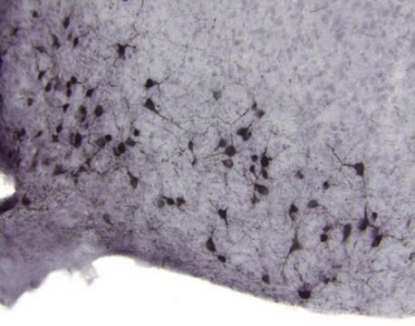
immunohistochemistry (formalin-fixed, paraffin-embedded sections): suitable
western blot: suitable

isotype

IgG1κ

NCBI accession no.

UniProt accession no.

shipped in

wet ice

target post-translational modification

unmodified

Gene Information

human ... CHGA(1113)

General description

Chromogranin A (a protein of 439 amino acids which is encoded on chromosome 14) is present in neuroendocrine cells throughout the body, including the neuroendocrine cells of the large and small intestine, adrenal medulla and pancreatic islets. It is an excellent marker for carcinoid tumors, phenochromocytomas, paragangliomas, and other neuroendocrine tumors. Coexpression of chromogranin A and neuron specific enolase (NSE) is common in neuroendocrine neoplasms.

Specificity

Chromogranin, an acidic protein having a molecular weight of about 68 kDa abides in the polypeptide-hormone-containing secretory granules of human endocrine tissues. Anti-chromogranin A can be used to stain most normal and neoplastic endocrine tissues containing secretory granules (exceptions include cells from the hypothalamus and posterior pituitary, which are known to contain secretory granules). The amount of staining is usually proportional to the number of secretory granules present (1).

Application

Detect Chromogranin A using this Anti-Chromogranin A Antibody, clone LK2H10 validated for use in IH, IH(P) & WB.
Immunohistochemistry:
A 1-10 μg/mL concentration of a previous lot was used in IH.

Optimal working dilutions must be determined by end user.

Packaging

500 μL

Quality

Routinely evaluated by Western Blot on Mouse Pancreas lysates.

Western Blot Analysis:
1:1000 dilution of this lot detected Chromogranin A on 10 μg of Mouse Pancreas lysate.

Target description

50 kDa

Linkage

Replaces: CBL219

Physical form

Format: Purified
Purified mouse monoclonal IgG1 in buffer containing 0.05M Borate buffered saline, 0.15M NaCl, pH 8.2 with 0.1% sodium azide.

Analysis Note

Control
Pancreas, adrenal gland, bowel and thyroid tissues Immunoblot: PC12 cell lysate, human pancreatic tissue

Other Notes

Concentration: Please refer to the Certificate of Analysis for the lot-specific concentration.

Legal Information

CHEMICON is a registered trademark of Merck KGaA, Darmstadt, Germany

Optional

pictograms

Health hazard

signalword

Danger

hcodes

Hazard Classifications

Repr. 1B

Storage Class

6.1D - Non-combustible acute toxic Cat.3 / toxic hazardous materials or hazardous materials causing chronic effects

wgk_germany

nwg

flash_point_f

Not applicable

flash_point_c

Not applicable


Certificados de análisis (COA)

Busque Certificados de análisis (COA) introduciendo el número de lote del producto. Los números de lote se encuentran en la etiqueta del producto después de las palabras «Lot» o «Batch»

¿Ya tiene este producto?

Encuentre la documentación para los productos que ha comprado recientemente en la Biblioteca de documentos.

Visite la Librería de documentos

Specific endocrine tissue marker defined by a monoclonal antibody.
Lloyd, R V and Wilson, B S
Science (New York, N.Y.), 222, 628-630 (1983)
Fausto J Rodriguez et al.
Brain pathology (Zurich, Switzerland), 19(4), 623-629 (2008-08-20)
Co-deletion of chromosome arms 1p and 19q, characteristic of oligodendroglial tumors, was recently found to be mediated by t(1;19)(q10;p10). To evaluate the prevalence of 1p19q co-deletion and t(1;19) in extraventricular neurocytomas (EVN), we studied tumors from 23 patients, including 13
Tieying Hou et al.
Cancer cytopathology, 128(10), 725-732 (2020-06-24)
Insulinoma-associated protein 1 (INSM1) has recently emerged as a reliable nuclear immunostaining marker for detecting neuroendocrine tumors (NETs) in paraffin-embedded surgical samples and cytologic cell blocks, but the reliability of INSM1 staining on cytologic smears is understudied. This study investigated
Neuron precursor features of spindle cell oncocytoma of adenohypophysis.
Sanda Alexandrescu,Robert E Brown,Nitin Tandon,Meenakshi B Bhattacharjee
Annals of Clinical and Laboratory Science null
Noèlia Téllez et al.
American journal of physiology. Endocrinology and metabolism, 311(3), E554-E563 (2016-07-14)
Limitations in β-cell regeneration potential in middle-aged animals could contribute to the increased risk to develop diabetes associated with aging. We investigated β-cell regeneration of middle-aged Wistar rats in response to two different regenerative stimuli: partial pancreatectomy (Px + V)

Nuestro equipo de científicos tiene experiencia en todas las áreas de investigación: Ciencias de la vida, Ciencia de los materiales, Síntesis química, Cromatografía, Analítica y muchas otras.

Póngase en contacto con el Servicio técnico