Saltar al contenido
Merck
Todas las fotos(2)

Key Documents

16-256

Sigma-Aldrich

Anti-Phosphatidylserine Antibody, clone 1H6, Alexa Fluor 488

clone 1H6, Upstate®, from mouse

Sinónimos:

488-Alexa Fluor Antibody, Alexa Fluor 488 Detection, Clone 1H6 Anti-Phosphatidylserine

Iniciar sesiónpara Ver la Fijación de precios por contrato y de la organización


About This Item

UNSPSC Code:
12352203
eCl@ss:
32160702
NACRES:
NA.41

biological source

mouse

Quality Level

conjugate

ALEXA FLUOR 488

antibody form

purified immunoglobulin

antibody product type

primary antibodies

clone

1H6, monoclonal

species reactivity

vertebrates

manufacturer/tradename

Upstate®

technique(s)

flow cytometry: suitable

isotype

IgG

shipped in

wet ice

target post-translational modification

phosphorylation (pSer)

General description

Significance:
Anti-Phosphatidylserine may be used to detect translocation of the membrane phospholipid phosphatidylserine (PS) from the inner to the outer cell membrane leaflet; it provides an alternative to Annexin V for quantitation of apoptosis.
Phosphatidylserine, or PS, is a naturally occurring, phospholipid nutrient. PS is essential to the functioning of all the cells of the body, but is most concentrated in the brain. Its relative abundance in this organ reflects its proven involvement in an assortment of nerve cell functions, including nerve transmitter release and synaptic activity. Clinical studies have suggested that PS can support brain functions that tend to decline with age.

Specificity

Recognizes phosphatidylserine (PS) in cell membranes.

Immunogen

Liposomes containing 70% phosphatidylserine and 30% phosphatidylglycerol.

Application

Detect Phosphatidylserine using this Anti-Phosphatidylserine Antibody, clone 1H6, Alexa Fluor 488 validated for use in FC.
Research Category
Apoptosis & Cancer
Research Sub Category
Apoptosis - Additional

Quality

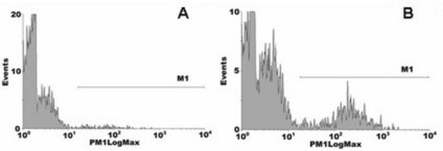
Flow Cytometry: 0.2 μg of this antibody detected Phosphatidylserine in fixed Jurkat cells (see page 2).

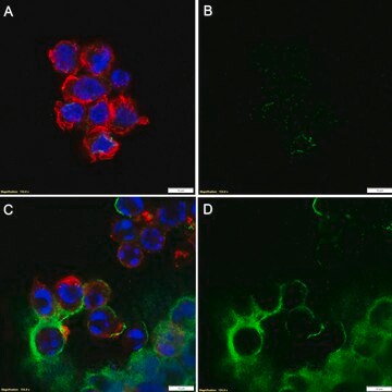
Apoptosis Assay: Time course for induction of apoptosis in Jurkat cells by staurosporine, measured using either an Anti-Phosphatidylserine, clone 1H6, Alexa Fluor 488 conjugate or an Annexin V FITC conjugate.

Physical form

Protein G Purified
Purified mouse monoclonal IgG in buffer containing PBS containing 1% BSA, 0.05% Tween, 0.05% sodium azide. Liquid at 4ºC.

Storage and Stability

Stable for 1 year at from date of receipt.

Analysis Note

Control
Negative Control: Catalog # 16-240, Normal Mouse IgG, Alexa Fluor 488-conjugate.

Other Notes

Concentration: Please refer to the Certificate of Analysis for the lot-specific concentration.

Legal Information

ALEXA FLUOR is a trademark of Life Technologies
UPSTATE is a registered trademark of Merck KGaA, Darmstadt, Germany

Disclaimer

Alexa Fluor is a registered trademark of Molecular Probes, Inc.

Unless otherwise stated in our catalog or other company documentation accompanying the product(s), our products are intended for research use only and are not to be used for any other purpose, which includes but is not limited to, unauthorized commercial uses, in vitro diagnostic uses, ex vivo or in vivo therapeutic uses or any type of consumption or application to humans or animals.

¿No encuentra el producto adecuado?  

Pruebe nuestro Herramienta de selección de productos.

Storage Class

12 - Non Combustible Liquids

wgk_germany

WGK 2

flash_point_f

Not applicable

flash_point_c

Not applicable


Certificados de análisis (COA)

Busque Certificados de análisis (COA) introduciendo el número de lote del producto. Los números de lote se encuentran en la etiqueta del producto después de las palabras «Lot» o «Batch»

¿Ya tiene este producto?

Encuentre la documentación para los productos que ha comprado recientemente en la Biblioteca de documentos.

Visite la Librería de documentos

Yuta Nakazawa et al.
eLife, 10 (2021-11-10)
Although tumor-infiltrating regulatory T (Treg) cells play a pivotal role in tumor immunity, how Treg cell activation are regulated in tumor microenvironments remains unclear. Here, we found that mice deficient in the inhibitory immunoreceptor CD300a on their dendritic cells (DCs)
Yi An et al.
Scientific reports, 11(1), 6392-6392 (2021-03-20)
Head and neck squamous cell carcinomas (HNSCC) induced by human papillomavirus (HPV) have increased recently in the US. However, the distinct alterations of molecules involved in the death pathways and drug effects targeting inhibitor of apoptosis proteins (IAPs) have not
Lazar Mandinov et al.
Proceedings of the National Academy of Sciences of the United States of America, 100(11), 6700-6705 (2003-05-20)
The induction of an acute inflammatory response followed by the release of polypeptide cytokines and growth factors from peripheral blood monocytes has been implicated in mediating the response to vascular injury. Because the Cu2+-binding proteins IL-1alpha and fibroblast growth factor
P Nusbaum et al.
Biochemical Society transactions, 32(Pt3), 477-479 (2004-05-26)
CD43 down-regulation during the apoptosis of PMN (polymorphonuclear cells) is not caused by proteolysis or internalization. Could it be released with bleb-derived membrane vesicles? Membrane blebbing was followed by microscopy on PMN 'synchronized' by an overnight incubation at 15 degrees
Mark C Weir et al.
PloS one, 12(7), e0181178-e0181178 (2017-07-21)
Acute myelogenous leukemia (AML) is often associated with activating mutations in the receptor tyrosine kinase, Flt3, including internal tandem duplications (ITDs) within the regulatory juxtamembrane region. Previous studies have linked Flt3-ITD to the activation of the Fes protein tyrosine kinase

Nuestro equipo de científicos tiene experiencia en todas las áreas de investigación: Ciencias de la vida, Ciencia de los materiales, Síntesis química, Cromatografía, Analítica y muchas otras.

Póngase en contacto con el Servicio técnico