Saltar al contenido
Merck
Todas las fotos(2)

Key Documents

05-390

Sigma-Aldrich

Anti-erbB-3/HER-3 Antibody, clone 2F12

clone 2F12, Upstate®, from mouse

Sinónimos:

Tyrosine kinase-type cell surface receptor HER3, lethal congenital contracture syndrome 2, v-erb-b2 avian erythroblastic leukemia viral oncogene homolog 3, v-erb-b2 erythroblastic leukemia viral oncogene homolog 3 (avian)

Iniciar sesiónpara Ver la Fijación de precios por contrato y de la organización


About This Item

UNSPSC Code:
12352203
eCl@ss:
32160702
NACRES:
NA.41

biological source

mouse

Quality Level

antibody form

purified antibody

antibody product type

primary antibodies

clone

2F12, monoclonal

species reactivity

human, bovine, mouse, rat

manufacturer/tradename

Upstate®

technique(s)

immunocytochemistry: suitable
immunohistochemistry: suitable
immunoprecipitation (IP): suitable
western blot: suitable

isotype

IgG2aκ

NCBI accession no.

UniProt accession no.

shipped in

wet ice

target post-translational modification

unmodified

Gene Information

human ... ERBB3(2065)

General description

The ErbB3 gene encodes a member of the epidermal growth factor receptor (EGFR) family of receptor tyrosine kinases. ErbB3 is a membrane-bound protein which has a neuregulin binding domain but not an active kinase domain. It can therefore bind this ligand but cannot convey a signal into the cell via protein phosphorylation. However it does form heterodimers with other EGF receptor family members which do have kinase activity. Heterodimerization leads to the activation of pathways which lead to cell proliferation or differentiation. Amplification of this gene and/or overexpression of its protein have been reported in numerous cancers including prostate, bladder and breast tumors. Alternate transcriptional splice variants encoding different isoforms have been characterized. One isoform lacks the intermembrane region and is secreted outside the cell. This form acts to modulate the activity of the membrane-bound form. Additional splice variants have also been reported but they have not been thoroughly characterized.

Specificity

Predicted to cross-react with mouse, rat, and cow.
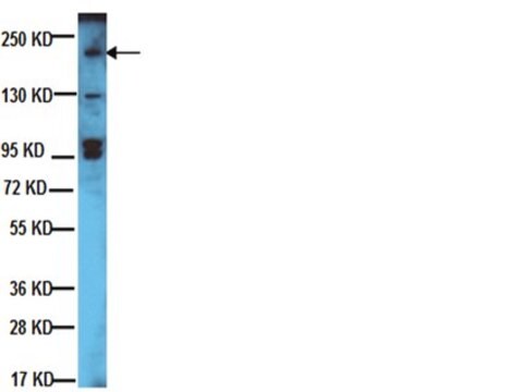
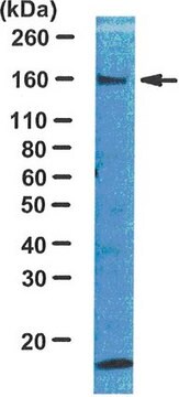
This antibody recognizes erbB-3/HER-3, Mr 185 kDa. Does not cross-react with erbB-1 (EGFR) and erbB-2/HER-2.

Immunogen

Epitope: a.a. 1295-1323 of human erbB-3
Recombinant rat erbB-3/HER-3 oncoprotein expressed in insect cells and purified by metal chelating chromatography. The epitope maps between amino acid residues 1295-1323 of human erbB-3. Clone 2F12.

Application

Detect erbB-3/HER-3 using this Anti-erbB-3/HER-3 Antibody, clone 2F12 validated for use in IC, IH, IP & WB.
Immunoprecipitation:
4 µg of a previous lot of this antibody immunoprecipitated erbB-3/HER-3 from human MDA and MCF-7 RIPA lysates.

Immunocytochemistry:
10 µg/mL of a previous lot of this antibody gave positive immunostaining for erbB-3/HER-3 in MCF-7cells fixed with 95% ethanol/5% acetic acid.

Immunohistochemistry:
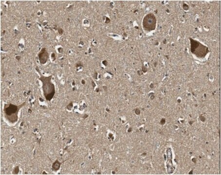
1:50 and 1:100 dilution of a previous lot detected erbB-3/HER-3 membrane staining in human normal and carcinoma breast tissue.
Research Category
Signaling
Research Sub Category
Growth Factors & Receptors

Quality

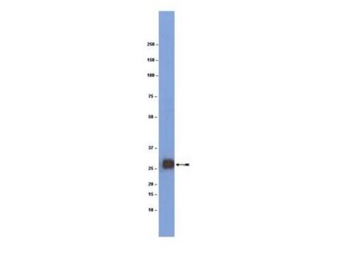
Routinely evaluated by Western Blot on RIPA lysates of human MCF-7 cells.

Western Blot Analysis:
0.1-2 µg/mL of this lot detected erbB-3/HER-3 in RIPA lysates of human MCF-7 cells. Previous lots have detected erbB-3/HER-3 in MDA RIPA lysates.

Target description

185 kDa

Physical form

Format: Purified
Protein A chromatography
Purified mouse monoclonal IgG2aκ in buffer containing 10 mM PBS, pH 7.4, 0.2% BSA, with 0.09% sodium azide. Frozen solution.

Storage and Stability

Stable for 1 year at -20°C from date of receipt.

Handling Recommendations:
Upon receipt, and prior to removing the cap, centrifuge the vial and gently mix the solution. Aliquot into microcentrifuge tubes and store at -20°C. Avoid repeated freeze/thaw cycles, which may damage IgG and affect product performance.

Analysis Note

Control
Human breast adenocarcinoma (well-differentiated), mouse brain lysate, MCF-7 cell lysate.

Other Notes

Concentration: Please refer to the Certificate of Analysis for the lot-specific concentration.

Legal Information

UPSTATE is a registered trademark of Merck KGaA, Darmstadt, Germany

Disclaimer

Unless otherwise stated in our catalog or other company documentation accompanying the product(s), our products are intended for research use only and are not to be used for any other purpose, which includes but is not limited to, unauthorized commercial uses, in vitro diagnostic uses, ex vivo or in vivo therapeutic uses or any type of consumption or application to humans or animals.

¿No encuentra el producto adecuado?  

Pruebe nuestro Herramienta de selección de productos.

Optional

Referencia del producto
Descripción
Precios

Storage Class

12 - Non Combustible Liquids

wgk_germany

WGK 2

flash_point_f

Not applicable

flash_point_c

Not applicable


Certificados de análisis (COA)

Busque Certificados de análisis (COA) introduciendo el número de lote del producto. Los números de lote se encuentran en la etiqueta del producto después de las palabras «Lot» o «Batch»

¿Ya tiene este producto?

Encuentre la documentación para los productos que ha comprado recientemente en la Biblioteca de documentos.

Visite la Librería de documentos

K M Darcy et al.
The journal of histochemistry and cytochemistry : official journal of the Histochemistry Society, 48(1), 63-80 (2000-02-01)
Studies were undertaken to examine the natural role of ErbB2, ErbB3, and ErbB4 during the development of normal rat mammary epithelial cells (MECs) in vivo and in vitro. Immunohistochemical analysis demonstrated that mammary gland terminal end buds expressed abundant ErbB2
Further evidence for the involvement of insulin receptor substrates in epidermal growth factor-induced activation of phosphatidylinositol 3-kinase.
T Fujioka,J H Kim,H Adachi,K Saito,M Tsujimoto,S Yokoyama,M Ui
European Journal of Biochemistry null
Abdelhamid Beji et al.
Clinical cancer research : an official journal of the American Association for Cancer Research, 18(4), 956-968 (2011-12-07)
Abnormal accumulation and dysregulation of the epidermal growth factor receptor family member HER3 is associated with the development of various human cancers including those of the breast, lung, and ovary. We have previously shown that in melanoma HER3 is frequently
Christèle Desbois-Mouthon et al.
Clinical cancer research : an official journal of the American Association for Cancer Research, 15(17), 5445-5456 (2009-08-27)
The insulin-like growth factor (IGF) signaling axis is frequently dysregulated in hepatocellular carcinoma (HCC). Therefore, we investigated whether the specific targeting of the IGF type 1 receptor (IGF-1R) might represent a new therapeutic approach for this tumor. Total and phosphorylated
Daniel J Zabransky et al.
Proceedings of the National Academy of Sciences of the United States of America, 112(45), E6205-E6214 (2015-10-29)
Recurrent human epidermal growth factor receptor 2 (HER2) missense mutations have been reported in human cancers. These mutations occur primarily in the absence of HER2 gene amplification such that most HER2-mutant tumors are classified as "negative" by FISH or immunohistochemistry

Nuestro equipo de científicos tiene experiencia en todas las áreas de investigación: Ciencias de la vida, Ciencia de los materiales, Síntesis química, Cromatografía, Analítica y muchas otras.

Póngase en contacto con el Servicio técnico