MABT818
Anti-Myosin-3 (MYH3) Antibody, clone BF-G6
clone BF-G6, from mouse
Synonym(s):
Myosin-3, Muscle embryonic myosin heavy chain, Myosin heavy chain 3, Myosin heavy chain, fast skeletal muscle, embryonic, SMHCE
About This Item
Recommended Products
biological source
mouse
Quality Level
antibody form
purified immunoglobulin
antibody product type
primary antibodies
clone
BF-G6, monoclonal
species reactivity
rat, human
species reactivity (predicted by homology)
bovine (based on 100% sequence homology)
technique(s)
ELISA: suitable
immunocytochemistry: suitable
immunofluorescence: suitable
immunohistochemistry: suitable
western blot: suitable
isotype
IgG1κ
NCBI accession no.
UniProt accession no.
shipped in
ambient
target post-translational modification
unmodified
Gene Information
human ... MYH3(4621)
General description
Specificity
Immunogen
Application
Cell Structure
ELISA Analysis: Clone BF-G6 hybridoma culture supernatant detected myosin from 10-week human fetus, but not myosin from 8-day new born muscle or adult skeletal muscle (Schiaffino, S., et al. (1986). Exp. Cell Res. 163(1):211-220).
Immunocytochemistry Analysis: Clone BF-G6 hybridoma culture supernatant immunostained aceton-fixed tumor cells in bone marrow aspiration from a child with rhobdomyosarcoma (Schiaffino, S., et al. (1986). Exp. Cell Res. 163(1):211-220).
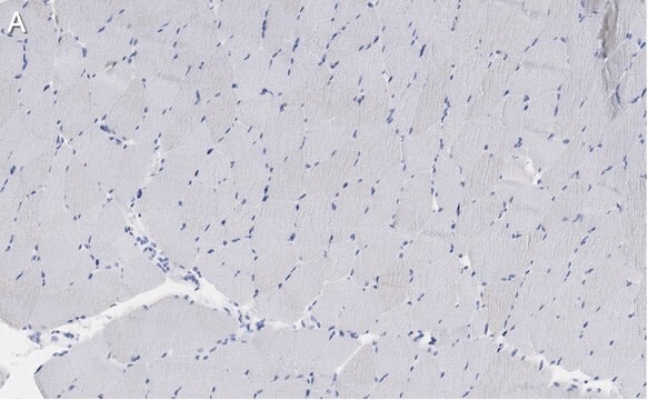
Immunofluorescence Analysis: Clone BF-G6 hybridoma culture supernatant immunostained serial transverse cryosections of rat extraocular (EO) and soleus muscles. BF-G6 staining pattern is similar, but not identical to that of MYH15 (Rossi, A.C., et al. (2010). J. Physiol. 588(Pt 2):353-364).
Immunofluorescence Analysis: Clone BF-G6 hybridoma culture supernatant immunostained frozen fetal muscle sections, while very few cells were stained in 8-week new born muscle tissue and no staining of 8-month infant muscle tissue was observed (Schiaffino, S., et al. (1986). Exp. Cell Res. 163(1):211-220).
Immunohistochemistry Analysis: Clone BF-G6 hybridoma culture supernatant immunostained tumor cells in frozen human rhobdomyosarcoma sections (Schiaffino, S., et al. (1986). Exp. Cell Res. 163(1):211-220).
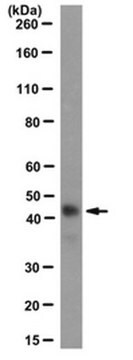
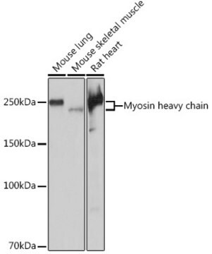
Western Blotting Analysis: Clone BF-G6 hybridoma culture supernatant detected myosin from 10-week human fetus, but not myosin from adult skeletal muscle (Schiaffino, S., et al. (1986). Exp. Cell Res. 163(1):211-220).
Quality
Isotyping Analysis: The identity of this monoclonal antibody is confirmed by isotyping test to be mouse IgG1κ.
Target description
Physical form
Storage and Stability
Other Notes
Disclaimer
Not finding the right product?
Try our Product Selector Tool.
Storage Class Code
12 - Non Combustible Liquids
WGK
WGK 1
Flash Point(F)
Not applicable
Flash Point(C)
Not applicable
Certificates of Analysis (COA)
Search for Certificates of Analysis (COA) by entering the products Lot/Batch Number. Lot and Batch Numbers can be found on a product’s label following the words ‘Lot’ or ‘Batch’.
Already Own This Product?
Find documentation for the products that you have recently purchased in the Document Library.
Our team of scientists has experience in all areas of research including Life Science, Material Science, Chemical Synthesis, Chromatography, Analytical and many others.
Contact Technical Service


