Skip to Content
Merck
All Photos(4)

Key Documents

HPA014561

Sigma-Aldrich

Anti-TMEM30A antibody produced in rabbit

Prestige Antibodies® Powered by Atlas Antibodies, affinity isolated antibody, buffered aqueous glycerol solution

Synonym(s):

Anti-C6orf67, Anti-CDC50A, Anti-FLJ10856, Anti-transmembrane protein 30A

Sign Into View Organizational & Contract Pricing


About This Item

UNSPSC Code:
12352203
Human Protein Atlas Number:
NACRES:
NA.43

biological source

rabbit

Quality Level

conjugate

unconjugated

antibody form

affinity isolated antibody

antibody product type

primary antibodies

clone

polyclonal

product line

Prestige Antibodies® Powered by Atlas Antibodies

form

buffered aqueous glycerol solution

species reactivity

human

technique(s)

immunohistochemistry: 1:50-1:200

immunogen sequence

VKSRDDSQLNGDSSALLNPSKECEPYRRNEDKPIAPCGAIANSMFNDTLELFLIGNDSYPIPIALKKKGIAWWTDKNVKFRNPPGGDNLEERFKGTTKPVNWLKPVYMLDSDPDNNGFI

UniProt accession no.

shipped in

wet ice

storage temp.

−20°C

target post-translational modification

unmodified

Gene Information

human ... TMEM30A(55754)

General description

TMEM30A (transmembrane protein 30A) is a functional homolog of Saccharomyces cerevisiae Lem3p protein. It has the conserved sequence QNHRRYVKS and is a transmembrane protein. The corresponding mRNA has a wide range of tissue expression. In cells expressing high level of TMEM30A, a major portion of this protein localizes to organelles such as, mitochondria, endoplasmic reticulum (ER) and Golgi bodies. This gene is localized to human chromosome 6q14.1, and encodes a protein composed of 361 amino acids. TMEM30A is also called CDC50A (cell division cycle protein 50A) and is a member of CDC50 family, which in humans includes- CDC50A, CDC50B and CDC50C.

Immunogen

transmembrane protein 30A recombinant protein epitope signature tag (PrEST)

Application

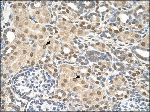
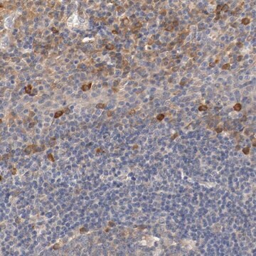
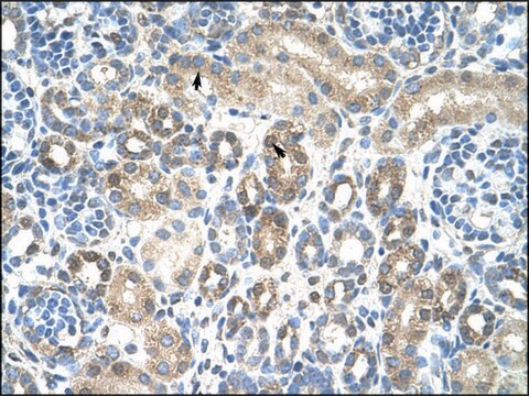
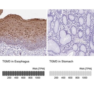
All Prestige Antibodies Powered by Atlas Antibodies are developed and validated by the Human Protein Atlas (HPA) project and as a result, are supported by the most extensive characterization in the industry.

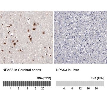
The Human Protein Atlas project can be subdivided into three efforts: Human Tissue Atlas, Cancer Atlas, and Human Cell Atlas. The antibodies that have been generated in support of the Tissue and Cancer Atlas projects have been tested by immunohistochemistry against hundreds of normal and disease tissues and through the recent efforts of the Human Cell Atlas project, many have been characterized by immunofluorescence to map the human proteome not only at the tissue level but now at the subcellular level. These images and the collection of this vast data set can be viewed on the Human Protein Atlas (HPA) site by clicking on the Image Gallery link. We also provide Prestige Antibodies® protocols and other useful information.

Biochem/physiol Actions

TMEM30A (transmembrane protein 30A) forms a heterodimeric complex with ATP8B1 P-type ATPase, and has a role in phospholipid import system. Extracellular choline phospholipids with short sn-2 residues act as substrates for this protein. It also forms a phospholipid flippase complex with ATP8A1, and facilitates the transfer of phosphatidylethanolamine to the cell membrane. This complex also enhances cell migration by facilitating the formation of membrane ruffles. Under-expression of TMEM30A leads to the exposure of phosphatidylserine on the outer leaflet of cell membrane, which acts as a signal to macrophages to phagocytose the particular cell. It also facilitates the uptake of antitumor drug perifosine by human cancer cells, most probably by association with P4-ATPase. It is a putative chaperone or β-subunit for ATP8B1 in hepatocytes. It also plays a role in the transport of ATP8B1 to plasma membrane, and thus, might be linked with ATP8B1-related disorders.

Features and Benefits

Prestige Antibodies® are highly characterized and extensively validated antibodies with the added benefit of all available characterization data for each target being accessible via the Human Protein Atlas portal linked just below the product name at the top of this page. The uniqueness and low cross-reactivity of the Prestige Antibodies® to other proteins are due to a thorough selection of antigen regions, affinity purification, and stringent selection. Prestige antigen controls are available for every corresponding Prestige Antibody and can be found in the linkage section.

Every Prestige Antibody is tested in the following ways:
  • IHC tissue array of 44 normal human tissues and 20 of the most common cancer type tissues.
  • Protein array of 364 human recombinant protein fragments.

Linkage

Corresponding Antigen APREST72841

Physical form

Solution in phosphate-buffered saline, pH 7.2, containing 40% glycerol and 0.02% sodium azide.

Legal Information

Prestige Antibodies is a registered trademark of Merck KGaA, Darmstadt, Germany

Disclaimer

Unless otherwise stated in our catalog or other company documentation accompanying the product(s), our products are intended for research use only and are not to be used for any other purpose, which includes but is not limited to, unauthorized commercial uses, in vitro diagnostic uses, ex vivo or in vivo therapeutic uses or any type of consumption or application to humans or animals.

Not finding the right product?  

Try our Product Selector Tool.

Storage Class Code

10 - Combustible liquids

WGK

WGK 1

Flash Point(F)

Not applicable

Flash Point(C)

Not applicable


Certificates of Analysis (COA)

Search for Certificates of Analysis (COA) by entering the products Lot/Batch Number. Lot and Batch Numbers can be found on a product’s label following the words ‘Lot’ or ‘Batch’.

Already Own This Product?

Find documentation for the products that you have recently purchased in the Document Library.

Visit the Document Library

Yuriko Katoh et al.
Oncology reports, 12(4), 939-943 (2004-09-18)
Bni1p, implicated in cell polarity control and microtubule regulation during yeast budding, is the Saccharomyces cerevisiae homolog of human Formin-homology proteins, such as FMN1, FMN2, FHOD1, FHOD3, FHDC1, GRID2IP, FMNL1, FMNL2, FMNL3, DIAPH1, DIAPH2, DIAPH3, DAAM1 and DAAM2. Cdc50p is
Rui Chen et al.
Journal of immunology (Baltimore, Md. : 1950), 186(5), 3215-3225 (2011-02-04)
Antitumor alkylphospholipids initiate apoptosis in transformed HL-60 and Jurkat cells while sparing their progenitors. 1-O-Alkyl-2-carboxymethyl-sn-glycero-3-phosphocholine (Edelfosine) like other short-chained phospholipids--inflammatory platelet-activating factor (PAF) and apoptotic oxidatively truncated phospholipids--are proposed to have intracellular sites of action, yet a conduit for these
Katsumori Segawa et al.
Science (New York, N.Y.), 344(6188), 1164-1168 (2014-06-07)
Phospholipids are asymmetrically distributed in the plasma membrane. This asymmetrical distribution is disrupted during apoptosis, exposing phosphatidylserine (PtdSer) on the cell surface. Using a haploid genetic screen in human cells, we found that ATP11C (adenosine triphosphatase type 11C) and CDC50A
Coen C Paulusma et al.
Hepatology (Baltimore, Md.), 47(1), 268-278 (2007-10-24)
Mutations in ATP8B1 cause progressive familial intrahepatic cholestasis type 1 and benign recurrent intrahepatic cholestasis type 1. Previously, we have shown in mice that Atp8b1 deficiency leads to enhanced biliary excretion of phosphatidylserine, and we hypothesized that ATP8B1 is a
Francisco Muñoz-Martínez et al.
Biochemical pharmacology, 80(6), 793-800 (2010-06-01)
Functional aminophospholipid translocases are composed of at least two proteins: an alpha subunit from the P4 subfamily of P-type ATPases and a beta subunit from the CDC50-Lem3p family. Over-expression and knockdown of the human beta subunit CDC50A in KB cells

Our team of scientists has experience in all areas of research including Life Science, Material Science, Chemical Synthesis, Chromatography, Analytical and many others.

Contact Technical Service