05-1003
Anti-phospho-Akt (Ser473) Antibody, clone 6F5
clone 6F5, from mouse
Synonym(s):
Protein kinase B, RAC-alpha serine/threonine-protein kinase, murine thymoma viral (v-akt) oncogene homolog-1, rac protein kinase alpha, v-akt murine thymoma viral oncogene homolog 1
About This Item
Recommended Products
biological source
mouse
Quality Level
antibody form
purified antibody
antibody product type
primary antibodies
clone
6F5, monoclonal
species reactivity
rat, mouse, vertebrates, human
technique(s)
ELISA: suitable
flow cytometry: suitable
immunofluorescence: suitable
immunohistochemistry: suitable
western blot: suitable
isotype
IgG1κ
NCBI accession no.
UniProt accession no.
shipped in
wet ice
target post-translational modification
phosphorylation (pSer473)
Gene Information
human ... AKT1(207)
rat ... Akt1S1(292887)
General description
Specificity
Immunogen
Application
EGF-treated A431 cells were fixed, permeabilized, and stained with Anti-phospho-Akt (Ser473), clone 6F5 (2 μg/mL) (red). Cy3-conjugated anti-mouse was used for detection and Phalloidin-AlexaFluor488 (green) was used to stain actin.
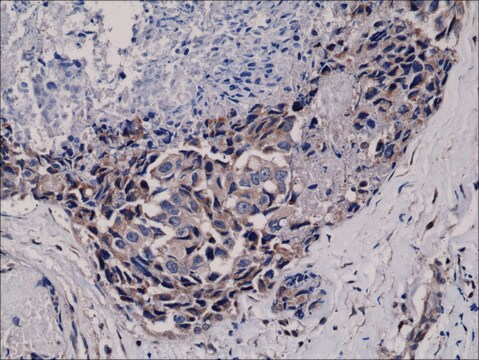
Immunohistochemistry:
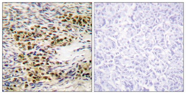
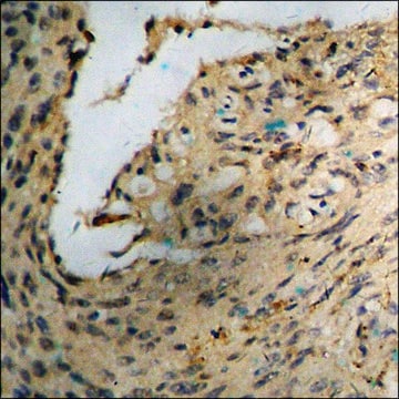
Anti-phospho-Akt (Ser473), clone 6F5 was diluted to 1:200, IHC-Select Detection with HRP-DAB.
Flow Cytometry:
Anti-phospho-Akt (Ser473) was used at a 1:800 dilution for this assay.
Quality
0.5-1 μg/mL of this lot detected phosphorylated Akt in lysates from mouse NIH-3T3 fibroblasts treated with 100 ng/mL PDGF for 20 minutes.
Target description
Physical form
Other Notes
Not finding the right product?
Try our Product Selector Tool.
recommended
Storage Class Code
12 - Non Combustible Liquids
WGK
WGK 1
Flash Point(F)
Not applicable
Flash Point(C)
Not applicable
Certificates of Analysis (COA)
Search for Certificates of Analysis (COA) by entering the products Lot/Batch Number. Lot and Batch Numbers can be found on a product’s label following the words ‘Lot’ or ‘Batch’.
Already Own This Product?
Find documentation for the products that you have recently purchased in the Document Library.
Articles
Autophagy is a highly regulated process that is involved in cell growth, development, and death. In autophagy cells destroy their own cytoplasmic components in a very systematic manner and recycle them.
Our team of scientists has experience in all areas of research including Life Science, Material Science, Chemical Synthesis, Chromatography, Analytical and many others.
Contact Technical Service