Skip to Content
Merck
All Photos(4)

Key Documents

05-593

Sigma-Aldrich

Anti-α-Dystroglycan Antibody, clone IIH6C4

ascites fluid, clone IIH6C4, Upstate®

Synonym(s):

Dystrophin-associated glycoprotein 1, dystroglycan 1, dystroglycan 1 (dystrophin-associated glycoprotein 1), dystrophin-associated glycoprotein-1, LARGE-glycan, Large glycan

Sign Into View Organizational & Contract Pricing


About This Item

UNSPSC Code:
12352203
eCl@ss:
32160702
NACRES:
NA.41

biological source

mouse

antibody form

ascites fluid

clone

IIH6C4, monoclonal

species reactivity

human, mouse, canine, rat, guinea pig, rabbit

manufacturer/tradename

Upstate®

technique(s)

immunofluorescence: suitable
immunohistochemistry: suitable
inhibition assay: suitable
western blot: suitable

isotype

IgM

NCBI accession no.

UniProt accession no.

shipped in

dry ice

target post-translational modification

unmodified

Gene Information

human ... DAG1(1605)

General description

Dystroglycans are essential elements of the neuromuscular junction (NMJ). The gene for dystroglycan is expressed as a precursor protein that is post translationally cleaved into a 156 kDa extracellular peripheral membrane protein called alpha dystroglycan and a 43 kDa transmembrane protein, beta dystroglycan. The latter protein contains a PPxY motif that promotes binding to WW domain containing proteins, such as utrophin and dystrophin. Phosphorylation at tyrosine 892 within the PPxY motif may regulate c Src interactions with beta dystroglycan, as well as inhibit interactions with WW domain proteins. In skeletal muscle, beta dystroglycan is normally localized to the plasma membrane, however phosphorylation of Tyr892 leads to localization of beta dystroglycan to endosomal compartments along with c Src. Thus, phosphorylation at Tyr892 may have important roles in altering the localization of beta dystroglycan during NMJ formation.
This product may be used for research purposes only. Diagnostic use of this product requires a license from the University of Iowa Research Foundation, 214 Technology Innovation Center, Iowa City, IA 52242

Specificity

This antibody recognizes α-Dystroglycan/LARGE-glycan, Mr 156 kDa.

Immunogen

Rabbit skeletal muscle membrane preparation. Clone IIH6C4.

Application

Inhibition of Laminin Binding to Dystroglycan: An independent lab has shown, in a nitrocellulose overlay experiment, that this antibody inhibits binding of 125I-laminin to dystroglycan (Ervasti, J., et al. (1993).

Western Blot Analysis: A previous lot of this antibody was used on mouse muscle tissue lysate 3 months after shRNA induction, and in littermate controls (ctrl) and LARGE-null negative control (myd) (Goddeeris, M., et al. 2013, Nature).

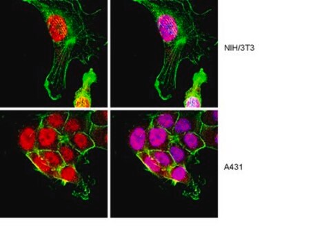
Immunofluorescence: A previous lot of this antibody was used to detect α-Dystroglycan /LARGE-glycan in mouse muscle tissue (Goddeeris, M., et al. 2013, Nature).
Research Category
Metabolism
Research Sub Category
Muscle Physiology
This Anti-α-Dystroglycan Antibody, clone IIH6C4 is validated for use in IH, FUNC, WB for the detection of α-Dystroglycan.

Quality

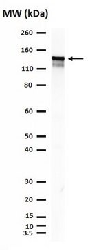
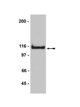
Routinely evaluated by western blot on rabbit skeletal muscle.

Western Blot Analysis:
A 1:1000-1:2000 dilution of this lot detected α-Dystroglycan/LARGE-glycan in rabbit skeletal muscle.
Note: The use of WGA purified protein results in significantly cleaner blots and immunoprecipitates.
Post-translational modification of dystroglycan causes band broadening.

Target description

156 kDa

Physical form

Mouse ascites, in PBS containing 0.05% sodium azide and 30% glycerol.
Liquid at -20ºC.
Unpurified

Storage and Stability

Stable for 1 year at -20°C from date of receipt. For maximum recovery of product, centrifuge the vial prior to removing the cap.

Analysis Note

Control
Rabbit skeletal muscle lysate.

Other Notes

Concentration: Please refer to the Certificate of Analysis for the lot-specific concentration.

Legal Information

UPSTATE is a registered trademark of Merck KGaA, Darmstadt, Germany

Disclaimer

Unless otherwise stated in our catalog or other company documentation accompanying the product(s), our products are intended for research use only and are not to be used for any other purpose, which includes but is not limited to, unauthorized commercial uses, in vitro diagnostic uses, ex vivo or in vivo therapeutic uses or any type of consumption or application to humans or animals.

Storage Class Code

12 - Non Combustible Liquids

WGK

WGK 2

Flash Point(F)

Not applicable

Flash Point(C)

Not applicable


Certificates of Analysis (COA)

Search for Certificates of Analysis (COA) by entering the products Lot/Batch Number. Lot and Batch Numbers can be found on a product’s label following the words ‘Lot’ or ‘Batch’.

Already Own This Product?

Find documentation for the products that you have recently purchased in the Document Library.

Visit the Document Library

Mice Lacking Dystrophin or {alpha} Sarcoglycan Spontaneously Develop Embryonal Rhabdomyosarcoma with Cancer-Associated p53 Mutations and Alternatively Spliced or Mutant Mdm2 Transcripts.
Fernandez K, Serinagaoglu Y, Hammond S, Martin LT, Martin PT
The American Journal of Pathology null
A role for the dystrophin-glycoprotein complex as a transmembrane linker between laminin and actin.
Ervasti, J M and Campbell, K P
The Journal of cell biology, 122, 809-823 (1993)
Susan Sparks et al.
BMC neurology, 7, 3-3 (2007-01-31)
Hereditary Inclusion Body Myopathy (HIBM) is an autosomal recessive, adult onset, non-inflammatory neuromuscular disorder with no effective treatment. The causative gene, GNE, codes for UDP-N-acetylglucosamine 2-epimerase/N-acetylmannosamine kinase, which catalyzes the first two reactions in the synthesis of sialic acid. Reduced
Francesca Sciandra et al.
BMC research notes, 10(1), 601-601 (2017-11-22)
Dystroglycan (DG) is an adhesion complex formed by two subunits, α-DG and β-DG. In skeletal muscle, DG is part of the dystrophin-glycoprotein complex that is crucial for sarcolemma stability and it is involved in a plethora of muscular dystrophy phenotypes.
B Wu et al.
Gene therapy, 21(9), 785-793 (2014-06-20)
Antisense therapy with both chemistries of phosphorodiamidate morpholino oligomers (PMOs) and 2'-O-methyl phosphorothioate has demonstrated the capability to induce dystrophin expression in Duchenne muscular dystrophy (DMD) patients in phase II-III clinical trials with benefit in muscle functions. However, potential of

Our team of scientists has experience in all areas of research including Life Science, Material Science, Chemical Synthesis, Chromatography, Analytical and many others.

Contact Technical Service