Performing a Purification of IgG Antibodies with Protein G Sepharose® 4 Fast Flow
`
Extracted from Affinity Chromatography Vol. 1: Antibodies, GE Healthcare, 2016
Extracted from Affinity Chromatography Vol. 1: Antibodies, GE Healthcare, 2016
Protein G Sepharose® 4 Fast Flow consists of 90 µm beads of highly cross-linked agarose, which provide a robust and stable chromatography matrix that allows high flow rates. The medium is a good choice for general-purpose capture of antibodies and scale-up in the laboratory.
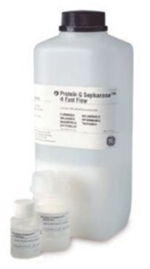
Protein G Sepharose® 4 Fast Flow is available as a bulk medium (Figure 3.2) for packing in XK, HiScale, and Tricorn columns (Figure 3.3) at laboratory scale. Furthermore, Protein G Sepharose® 4 Fast Flow can be packed in larger columns for industrial-scale purification of monoclonal and polyclonal antibodies.

Figure 3.2.Protein G Sepharose® 4 Fast Flow is available in various bulk (lab packs) sizes for laboratory and process-scale applications.
Column packing
Refer to Column packing and preparation for general column packing guidelines.
Sepharose® 4 Fast Flow chromatography media should be packed in XK or Tricorn columns in a two-step procedure: Do not exceed 0.3 bar (0.03 MPa) in the first step and 3.5 bar (0.35 MPa) in the second step. If the packing equipment does not include a pressure gauge, use a packing flow rate of 2.5 mL/min (XK 16/20 column) or 1.0 mL/min (Tricorn 10/100 column) in the first step, and 14 mL/min (XK 16/20 column) or 5.5 mL/min (Tricorn 10/100 column) in the second step.

Figure 3.3.Empty Tricorn columns for packing. Once packed, these columns can be used for purification using either a pump or chromatography system.
- Equilibrate all material to the temperature at which the purification will be performed and de-gas the medium slurry.
- Eliminate air from the column end-piece and adapter by flushing the end pieces with binding buffer. Make sure no air has been trapped under the column bed support. Close the column leaving the bed support covered with distilled water.
- Resuspend the medium and pour the slurry into the column in a single, continuous motion. Pouring the slurry down a glass rod held against the wall of the column minimizes the formation of air bubbles.
- If using a packing reservoir, immediately fill the remainder of the column with distilled water. Mount the adapter or lid of the packing reservoir and connect the column to a pump. Avoid trapping air bubbles under the adapter or in the inlet tubing.
- Open the bottom outlet of the column and set the pump to run at the desired flow rate.
- Maintain packing flow rate for at least 3 bed volumes after a constant bed height is reached. Mark the bed height on the column.
- Stop the pump and close the column outlet.
- If using a packing reservoir, disconnect the reservoir and fit the adapter to the column.
- With the adapter inlet disconnected, push the adapter down into the column until it reaches the mark. Allow the packing solution to flush the adapter inlet. Lock the adapter in position.
- Connect the column to a pump or a chromatography system and start equilibration. Re-adjust the adapter if necessary.
For subsequent chromatography procedures, do not exceed 75% of the packing flow rate.
Sample preparation
Centrifuge samples (10 000 × g for 10 min) to remove cells and debris. Filter through a 0.45 µM filter.
The sample should have a pH around 7.0 before applying to the column. If required, adjust sample conditions to the pH and ionic strength of the binding buffer by either buffer exchange on a desalting column (Desalting and buffer exchange) or dilution and pH adjustment.
Buffer preparation
Binding buffer: 0.02 M sodium phosphate, pH 7.0
Elution buffer: 0.1 M glycine-HCl, pH 2.7
Neutralizing buffer: 1 M Tris-HCl, pH 9.0
Water and chemicals used for buffer preparation should be of high purity.
Filter buffers through a 0.45 µM filter before use.
Purification
- Prepare collection tubes by adding 60 to 200 µL of 1 M Tris-HCl, pH 9.0 per milliliter of fraction to be collected.
To preserve the activity of acid-labile IgG, we recommend adding 60 to 200 µL of 1 M Tris-HCl pH 9.0 to collection tubes, which ensures that the final pH of the sample will be approximately neutral.
- If the column contains 20% ethanol, wash it with 5 column volumes of distilled water. Use a linear flow rate of 50 to 100 cm/h.
Appendix 7 for information on how to convert linear flow (cm/h) to volumetric flow rates (mL/min).
- Equilibrate the column with 5 to 10 column volumes of binding buffer at a linear flow rate of 150 cm/h.
- Apply the pretreated sample.
- Wash with binding buffer until the absorbance reaches the baseline.
- Elute with elution buffer using a step or linear gradient. For step elution, 5 column volumes of elution buffer are usually sufficient. For linear gradient elution, a shallow gradient over 20 column volumes allows separation of proteins with similar binding strengths.
- After elution, regenerate the column by washing it with 5 to 10 column volumes of binding buffer. The column is now ready for a new purification.
Desalt and/or transfer purified IgG fractions to a suitable buffer using a desalting column (Desalting and buffer exchange).
Storage
Store in 20% ethanol at 2 °C to 8 °C.
如要继续阅读,请登录或创建帐户。
暂无帐户?