推荐产品
生物源
mouse
品質等級
抗體表格
purified antibody
抗體產品種類
primary antibodies
無性繁殖
P4H2, monoclonal
物種活性
human
技術
immunocytochemistry: suitable
western blot: suitable
同型
IgG1κ
NCBI登錄號
UniProt登錄號
運輸包裝
ambient
目標翻譯後修改
unmodified
基因資訊
human ... DUX4(100288687)
一般說明
Double homeobox protein 4 (UniProt Q9UBX2; also known as Double homeobox protein 10) is encoded by the DUX4 (also known as DUX10) gene (Gene ID 100288687) in human. DUX4 is a double homeobox transcription factor that is normally expressed in the testis. Contraction or hypomethylation of D4Z4 macrosatellite repeats on chromosome 4q35 causes aberrant DUX4 expression in skeletal muscle, a hallmark of the incurable disease facioscapulohumeral muscular dystrophy (FSHD), characterized by skeletal muscle weakness and wasting. Gene expression data analysis identifies β-catenin as the main coordinator of FSHD-associated perturbation of protein interaction signallings, including those of canonical Wnt, HIF1-α and TNF-α. In addition, DUX4 expression causes proteolytic degradation of UPF1, a central component of the nonsense-mediated decay (NMD) machinery, and accumulation NMD substrate RNAs including DUX4 mRNA, such that NMD downregulation by DUX4 expression stabilizes DUX4 mRNA through a double-negative feedback loop in FSHD muscle cells. FSHD muscle expresses a low abundance DUX4 mRNA that produces the full-length DUX4 protein. In contrast to control skeletal muscle and most other somatic tissues, full-length DUX4 transcript and protein is expressed at relatively abundant levels in human testis, most likely in the germ-line cells. Induced pluripotent (iPS) cells also express full-length DUX4 and differentiation of iPS cells to embryoid bodies suppresses expression of full-length DUX4, whereas expression of full-length DUX4 persists in differentiated FSHD iPS cells.
特異性
Clone P4H2 is specific to DUX4 (UniProt Q9UBX2) and does not recognize DUX4c (DUX4L9; UniProt Q6RFH8) or the spliced DUX4 short isoform DUX4-s (UniProt E2JJS1) that lacks the C-terminal epitope region targeted by clone P4H2 (Geng, L.N., et al. (2011). Hybridoma (Larchmt). 30(2):125-130; Snider, L., et al. (2010). PLoS Genet. 6(10):e1001181).
免疫原
GST-tagged recombinant human DUX4 C-terminal fragment.
應用
Detect DUX4 using this mouse monoclonal Anti-DUX4 Antibody, clone P4H2, Cat. No. MABS1348, validated for use in Immunocytochemistry and Western Blotting.
Research Category
Signaling
Signaling
Western Blotting Analysis: 1 µg/mL from a representative lot detected DUX4 in 10 µg of human heart and ovary tissue lysates.
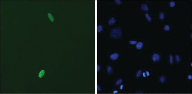
Immunocytochemistry Analysis: A representative lot detected full-length DUX4 (DUX4-FL) nuclear immunoreactivity at a higher frequency among differentiated CD56+ myogenic cells from facioscapulohumeral dystrophy (FSHD) than non-FSHD individuals (Jones, T.I., et al. (2012). Hum. Mol. Genet. 21(20):4419-4430).
Immunocytochemistry Analysis: A representative lot detected exogenously expressed full-length human DUX4 (DUX4-FL) in 2% paraformaldehyde-fixed and 1% Triton X-100-permeabilized C2C12 mouse myoblasts (Geng, L.N., et al. (2011). Hybridoma (Larchmt). 30(2):125-130).
Immunocytochemistry Analysis: A representative lot and another DUX4 antibody that targets an N-terminal region epitope co-stained full-length DUX4 (DUX4-FL) in the nucleus in ~0.1% of cultured facioscapulohumeral dystrophy (FSHD) muscle cells fixed with 2% paraformaldehyde and permeabilized with 1% Triton X-100 (Snider, L., et al. (2010). PLoS Genet. 6(10):e1001181).
Western Blotting Analysis: A representative lot detected exogenously expressed human DUX4, but not DUX4c, in transfected C2C12 mouse myoblasts. No target band was detected in lysate from untransfected cells (Geng, L.N., et al. (2011). Hybridoma (Larchmt). 30(2):125-130).
Western Blotting Analysis: A representative lot detected DUX4 immunoprecipitated from human testis tissue lysate by another DUX4 antibody that targets an N-terminal region epitope (Snider, L., et al. (2010). PLoS Genet. 6(10):e1001181).
品質
Evaluated by Western Blotting in human testis tissue lysate.
Western Blotting Analysis: 0.5 µg/mL of this antibody detected DUX4 in 10 µg of human testis tissue lysate.
Western Blotting Analysis: 0.5 µg/mL of this antibody detected DUX4 in 10 µg of human testis tissue lysate.
標靶描述
~60/45 kDa observed. 44.94 kDa calculated. The larger-than-calculated target band size is consistent with that reported in the literature (Geng, L.N., et al. (2011). Hybridoma (Larchmt). 30(2):125-130; Snider, L., et al. (2010). PLoS Genet. 6(10):e1001181). Uncharacterized bands may be observed in some lysate(s).
外觀
Protein G purified
Format: Purified
Purified mouse IgG1 in buffer containing 0.1 M Tris-Glycine (pH 7.4), 150 mM NaCl with 0.05% sodium azide.
儲存和穩定性
Stable for 1 year at 2-8°C from date of receipt.
其他說明
Concentration: Please refer to lot specific datasheet.
免責聲明
Unless otherwise stated in our catalog or other company documentation accompanying the product(s), our products are intended for research use only and are not to be used for any other purpose, which includes but is not limited to, unauthorized commercial uses, in vitro diagnostic uses, ex vivo or in vivo therapeutic uses or any type of consumption or application to humans or animals.
未找到合适的产品?
试试我们的产品选型工具.
儲存類別代碼
12 - Non Combustible Liquids
水污染物質分類(WGK)
WGK 1
閃點(°F)
Not applicable
閃點(°C)
Not applicable
我们的科学家团队拥有各种研究领域经验,包括生命科学、材料科学、化学合成、色谱、分析及许多其他领域.
联系技术服务部门