Skip to Content
Merck
All Photos(1)

Documents

MAB3230

Sigma-Aldrich

Anti-Cytokeratin 10 Antibody, clone RKSE60

clone RKSE60, Chemicon®, from mouse

Sign Into View Organizational & Contract Pricing


About This Item

UNSPSC Code:
12352203
eCl@ss:
32160702
NACRES:
NA.41

biological source

mouse

Quality Level

antibody form

purified immunoglobulin

antibody product type

primary antibodies

clone

RKSE60, monoclonal

species reactivity

rat, canine, human, mouse

manufacturer/tradename

Chemicon®

technique(s)

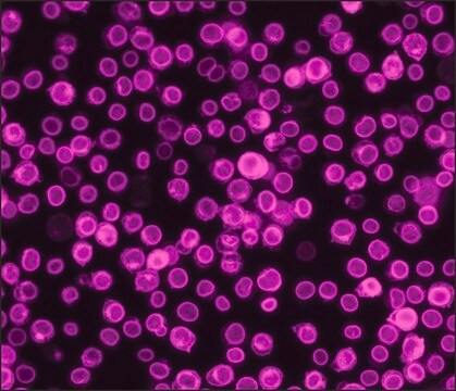
flow cytometry: suitable
immunocytochemistry: suitable
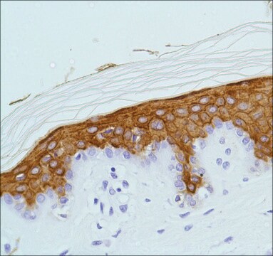
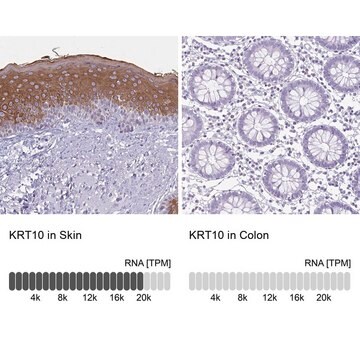
immunohistochemistry: suitable
western blot: suitable

isotype

IgG1

NCBI accession no.

UniProt accession no.

shipped in

dry ice

target post-translational modification

unmodified

Gene Information

human ... KRT10(3858)

General description

Cytokeratins are a subfamily of intermediate filament proteins and are characterized by a remarkable biochemical diversity, represented in epithelial tissues by at least 20 different polypeptides. They range in molecular weight from between 40 kDa and 68 kDa and isoelectric pH between 4.9 - 7.8, The individual cytokeratin polypeptides are designated 1-20. The various epithelia in the human body usually express cytokeratins which are not only characteristic of the type of epithelium, but also related to the degree of maturation or differentiation within an epithelium. Cytokeratin subtype expression patterns are used to an increasing extent in the distinction of different types of epithelial malignancies.

Specificity

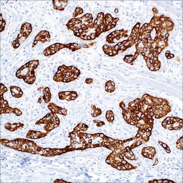
Cytokeratin 10. MAB3230 reacts with keratinizing stratified epithelia and in differentiated areas of highly differentiated squamous cell carcinomas. For the recognition of keratinizing cells in squamous cell carcinomas from epidermis, lung, bladder, cervix, esophagus etc. The antibody reacts only with suprabasal cells of the epidermis and not with basal cells.

Immunogen

Cytoskeletal preparation from human squamous keratinizing epithelium.

Application

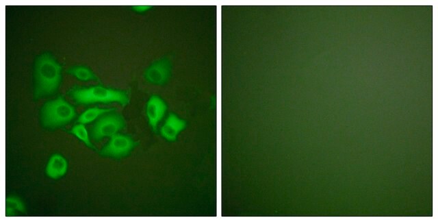
Anti-Cytokeratin 10 Antibody, clone RKSE60 detects level of Cytokeratin 10 & has been published & validated for use in FC, WB, IC, IH.
Research Category
Cell Structure
Research Sub Category
Cytokeratins
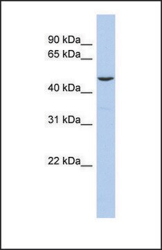
Western blot

Immunohistochemistry on frozen tissue sections.

Immunocytochemistry

Flow cytometry

Optimal working dilutions must be determined by the end user.

Physical form

Format: Purified
Liquid in buffer with 0.1% sodium azide.

Storage and Stability

Maintain at -20°C in undiluted aliquots up to 6 months. Avoid repeated freeze/thaw cycles.

Other Notes

Concentration: Please refer to the Certificate of Analysis for the lot-specific concentration.

Legal Information

CHEMICON is a registered trademark of Merck KGaA, Darmstadt, Germany

Disclaimer

Unless otherwise stated in our catalog or other company documentation accompanying the product(s), our products are intended for research use only and are not to be used for any other purpose, which includes but is not limited to, unauthorized commercial uses, in vitro diagnostic uses, ex vivo or in vivo therapeutic uses or any type of consumption or application to humans or animals.

Not finding the right product?  

Try our Product Selector Tool.

Storage Class Code

12 - Non Combustible Liquids

WGK

WGK 2

Flash Point(F)

Not applicable

Flash Point(C)

Not applicable


Certificates of Analysis (COA)

Search for Certificates of Analysis (COA) by entering the products Lot/Batch Number. Lot and Batch Numbers can be found on a product’s label following the words ‘Lot’ or ‘Batch’.

Already Own This Product?

Find documentation for the products that you have recently purchased in the Document Library.

Visit the Document Library

Loss of nucleoplasmic LAP2alpha-lamin A complexes causes erythroid and epidermal progenitor hyperproliferation.
Nana Naetar,Barbara Korbei,Serguei Kozlov,Marc A Kerenyi,Daniela Dorner,Rosana Kral et al.
Nature Cell Biology null
Atsushi Tokuriki et al.
Carcinogenesis, 30(9), 1645-1650 (2009-07-10)
Inhibitor of DNA binding 2 (Id2) is a negative regulator of basic helix-loop-helix transcription factors and is involved in the control of cellular differentiation and proliferation. By using a two-step chemical carcinogenesis protocol, we evaluated the role of Id2 in
Masashi Miyai et al.
The Journal of investigative dermatology, 136(9), 1848-1857 (2016-05-22)
Mammalian epidermis is a stratified epithelium composed of distinct layers of keratinocytes. The outermost cornified layer is a primary barrier that consists of a cornified envelope, an insoluble structure assembled by cross-linked scaffold proteins, and a surrounding mixture of lipids.
Inflammatory marker analysis in psoriatic skin under topical phosphodiesterase 4 inhibitor treatment.
Lennart M Roesner et al.
The Journal of allergy and clinical immunology, 140(4), 1184-1187 (2017-05-17)
Ryo Ichijo et al.
Nature communications, 8(1), 508-508 (2017-09-13)
The skin surface area varies flexibly in response to body shape changes. Skin homeostasis is maintained by stem cells residing in the basal layer of the interfollicular epidermis. However, how the interfollicular epidermal stem cells response to physiological body shape

Our team of scientists has experience in all areas of research including Life Science, Material Science, Chemical Synthesis, Chromatography, Analytical and many others.

Contact Technical Service