Skip to Content
Merck
All Photos(4)

Key Documents

05-591

Sigma-Aldrich

Anti-Akt/PKB Antibody, PH Domain, clone SKB1

clone SKB1, Upstate®, from mouse

Synonym(s):

Anti-AKT, Anti-PKB, Anti-PKB-ALPHA, Anti-PRKBA, Anti-RAC, Anti-RAC-ALPHA

Sign Into View Organizational & Contract Pricing


About This Item

UNSPSC Code:
12352203
eCl@ss:
32160702

biological source

mouse

Quality Level

antibody form

affinity purified immunoglobulin

antibody product type

primary antibodies

clone

SKB1, monoclonal

species reactivity

rat, human, mouse

packaging

antibody small pack of 25 μg

manufacturer/tradename

Upstate®

technique(s)

flow cytometry: suitable
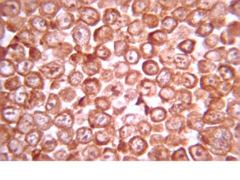
immunocytochemistry: suitable
immunoprecipitation (IP): suitable
western blot: suitable

isotype

IgG

NCBI accession no.

UniProt accession no.

shipped in

dry ice

target post-translational modification

unmodified

Gene Information

mouse ... Akt1(11651)

Related Categories

General description

Akt (protein kinase B), a serine/threonine kinase, has emerged as a critical enzyme in signal transduction pathways involved in cell proliferation, apoptosis, angiogenesis, and diabetes. In mammals three isoforms of Akt (α, β, γ or Akt 1, 2, 3) are reported that exhibit a high degree of homology, but differ slightly in the localization of their regulatory phosphorylation sites. Akt α is the predominant isoform in most tissues, whereas the highest expression of Akt β is observed in the insulin-responsive tissues, and Akt γ is abundant in brain tissue. Each Akt isoform is composed of three functionally distinct regions: an N-terminal pleckstrin homology (PH) domain that provides a lipid-binding module to direct Akt to PIP2 and PIP3, a central catalytic domain, and a C-terminal hydrophobic motif.

Application

Detect Akt/PKB using this Anti-Akt/PKB Antibody, PH Domain, clone SKB1 validated for use in FC, IC, IP & WB.

Quality

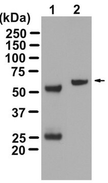
routinely evaluated by immunoblot on RIPA lysates from A431 cells

Target description

60 kDa

Physical form

Format: Purified
Protein G Purified immunoglobulin in Immunoaffinity Purified immunoglobulin in 0.02 M phosphate buffer, pH 7.6, 0.25M NaCl, 0.1% sodium azide and 30% glycerol.

Analysis Note

Control
Positive Antigen Control: Catalog #12-301, non-stimulated A431 cell lysate. Add 2.5µL of 2-mercaptoethanol/100µL of lysate and boil for 5 minutes to reduce the preparation. Load 20µg of reduced lysate per lane for minigels.

Other Notes

Concentration: Please refer to the Certificate of Analysis for the lot-specific concentration.

Legal Information

UPSTATE is a registered trademark of Merck KGaA, Darmstadt, Germany

Not finding the right product?  

Try our Product Selector Tool.


Certificates of Analysis (COA)

Search for Certificates of Analysis (COA) by entering the products Lot/Batch Number. Lot and Batch Numbers can be found on a product’s label following the words ‘Lot’ or ‘Batch’.

Already Own This Product?

Find documentation for the products that you have recently purchased in the Document Library.

Visit the Document Library

Evaluation of approaches to generation of tissue-specific knock-in mice.
Bayascas, JR; Sakamoto, K; Armit, L; Arthur, JS; Alessi, DR
The Journal of Biological Chemistry null
Regulation of AMP-activated protein kinase by multisite phosphorylation in response to agents that elevate cellular cAMP.
Hurley, RL; Barre, LK; Wood, SD; Anderson, KA; Kemp, BE; Means, AR; Witters, LA
The Journal of Biological Chemistry null
Important role of the LKB1-AMPK pathway in suppressing tumorigenesis in PTEN-deficient mice.
Huang X, Wullschleger S, Shpiro N, McGuire VA, Sakamoto K, Woods YL, McBurnie W et al.
The Biochemical Journal null
Heng-Liang Lin et al.
The Journal of pharmacology and experimental therapeutics, 323(1), 365-373 (2007-07-25)
Combretastatin A4 (CA4) is a drug that targets tumor vasculature to inhibit angiogenesis. Whether CA4 has a direct effect on gastric cancer is not known. We herein investigated the effect of CA4 on growth and metastasis of gastric cancer cells
HRT1 modulates vascular smooth muscle cell proliferation and apoptosis.
Wenli Wang, Chengyu Z Prince, Xing Hu, Matthew J Pollman
Biochemical and biophysical research communications null

Articles

Autophagy is a highly regulated process that is involved in cell growth, development, and death. In autophagy cells destroy their own cytoplasmic components in a very systematic manner and recycle them.

Our team of scientists has experience in all areas of research including Life Science, Material Science, Chemical Synthesis, Chromatography, Analytical and many others.

Contact Technical Service