Skip to Content
MilliporeSigma
All Photos(2)

Documents

SAB3500123

Sigma-Aldrich

Monoclonal Anti-PD-1 antibody produced in mouse

clone 12A7D7, purified immunoglobulin, buffered aqueous solution

Synonym(s):

Anti-CD279, Anti-PDCD-1, Anti-Programmed cell death 1

Sign Into View Organizational & Contract Pricing


About This Item

MDL number:
UNSPSC Code:
12352203
NACRES:
NA.41

biological source

mouse

conjugate

unconjugated

antibody form

purified immunoglobulin

antibody product type

primary antibodies

clone

12A7D7, monoclonal

form

buffered aqueous solution

species reactivity

rat, human, mouse

technique(s)

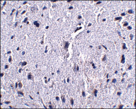
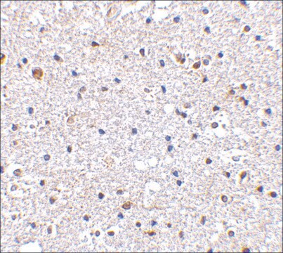
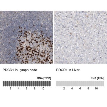
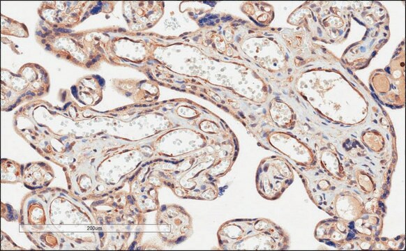
immunohistochemistry: suitable
indirect ELISA: suitable
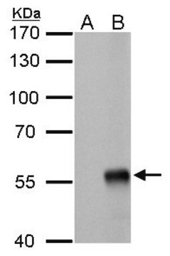
western blot: suitable

UniProt accession no.

shipped in

dry ice

storage temp.

−20°C

target post-translational modification

unmodified

Gene Information

human ... PDCD1(5133)

General description

Programmed death-1 (PD-1) is a type I transmembrane protein, encoded by the gene mapped to human chromosome 2q37.3. PD-1 belongs to the immunoglobulin receptor superfamily. The encoded protein is an immunoinhibitory receptor expressed by activated T cells, B cells, and myeloid cells. PD-1 is characterized with a single Ig-like variable (IgV) domain in the extracellular region and an immunoreceptor tyrosine-based inhibitory domain in the cytoplasmic region.

Immunogen

raised against an ~150 amino acid recombinant protein from near the terminus terminus of mouse PD-1.

Biochem/physiol Actions

Programmed death-1 (PD-1) gene is sensitive to systemic lupus erythematosus (SLE). Polymorphisms in PD-1 may participate in the growth of EOC (epithelial ovarian cancer). It acts as a negative regulator of T cells. It is also responsible for immune escape mediated by tumor cells. Interaction of PD-1 and PD-L1 (ligand of PD-1) results in inhibition of T cell receptor–mediated lymphocyte proliferation and cytokine secretion.

Features and Benefits

Evaluate our antibodies with complete peace of mind. If the antibody does not perform in your application, we will issue a full credit or replacement antibody. Learn more.

Target description

Cell-mediated immune responses are initiated by T lymphocytes that are themselves stimulated by cognate peptides bound to MHC molecules on antig en-presenting cells (APC). T-cell activation is generally self-limited as activated T cells express receptors such as PD-1 (also known as PDCD-1) that mediate inhibitory signals from the APC. PD-1 can bind two different but related ligands, PDL-1 and PDL-2. Upon binding to either of these ligands, signals generated by PD-1 inhibit the activation of the immune response in the absence of "danger signals" such as LPS or other molecules associated with bacteria or other pathogens. Evidence for this is seen in PD1-null mice who exhibit hyperactivated immune systems and autoimmune diseases. Despite its predicted molecular weight, PD-1 often migrates at higher molecular weight in SDS-PAGE.

Physical form

Supplied at approx. 1 mg/mL in phosphate buffered saline containing 0.02% sodium azide.

Disclaimer

Unless otherwise stated in our catalog or other company documentation accompanying the product(s), our products are intended for research use only and are not to be used for any other purpose, which includes but is not limited to, unauthorized commercial uses, in vitro diagnostic uses, ex vivo or in vivo therapeutic uses or any type of consumption or application to humans or animals.

Not finding the right product?  

Try our Product Selector Tool.

Storage Class

10 - Combustible liquids

wgk_germany

WGK 2

flash_point_f

Not applicable

flash_point_c

Not applicable


Certificates of Analysis (COA)

Search for Certificates of Analysis (COA) by entering the products Lot/Batch Number. Lot and Batch Numbers can be found on a product’s label following the words ‘Lot’ or ‘Batch’.

Already Own This Product?

Find documentation for the products that you have recently purchased in the Document Library.

Visit the Document Library

Christopher H Cogbill et al.
American journal of clinical pathology, 142(1), 88-98 (2014-06-14)
CD279 expression is used to help identify angioimmunoblastic T-cell lymphoma (AITL) or other T-cell lymphomas of T-follicular helper (TFH) cell origin; however, its utility in assessing lymphoid infiltrates in the bone marrow (BM) is not well established. Immunohistochemistry for CD279
Zhi-Chun Ding et al.
Cancer research, 74(13), 3441-3453 (2014-05-02)
In recent years, immune-based therapies have become an increasingly attractive treatment option for patients with cancer. Cancer immunotherapy is often used in combination with conventional chemotherapy for synergistic effects. The alkylating agent cyclophosphamide (CTX) has been included in various chemoimmunotherapy
Guoning Guo et al.
Diagnostic pathology, 9, 97-97 (2014-06-03)
Programmed death-1 (PD-1, Pdcd1)-deficient mice develop different types of autoimmune diseases depending on the mouse strain but its role in uterus development has not been reported. In this study, the expression of PD-1 and its ligands, PD-L1 and PD-L2, in
Alessandro Perrella et al.
Pathogens and disease, 72(2), 124-130 (2014-06-04)
We aimed to verify whether CD4(+)/CD25(+) T cells suppress CD4(+) T cells and secrete Granzyme B (GZB) during acute and chronic hepatitis C (CHC) infection. We enrolled 50 subjects: 20 patients with CHC (Group A), 15 healthy individuals (Group B)
Seon-Yeong Lee et al.
Arthritis & rheumatology (Hoboken, N.J.), 66(7), 1768-1778 (2014-03-20)
Intravenous immunoglobulin (IVIG) is used as a therapeutic agent in various autoimmune diseases. The aims of this study were to investigate the therapeutic effects of IVIG on collagen-induced arthritis (CIA) and identify the mechanism responsible for any therapeutic effects. IVIG

Our team of scientists has experience in all areas of research including Life Science, Material Science, Chemical Synthesis, Chromatography, Analytical and many others.

Contact Technical Service