Skip to Content
MilliporeSigma
All Photos(3)

Key Documents

ABN420

Sigma-Aldrich

Anti-RalDH2 (ALDH1A2)

from rabbit, purified by affinity chromatography

Synonym(s):

Retinal dehydrogenase 2, Aldehyde dehydrogenase family 1 member A2, RALDH 2, RalDH2, RALDH(II), Retinaldehyde-specific dehydrogenase type 2

Sign Into View Organizational & Contract Pricing


About This Item

UNSPSC Code:
12352203
eCl@ss:
32160702

biological source

rabbit

Quality Level

antibody form

affinity isolated antibody

antibody product type

primary antibodies

clone

polyclonal

purified by

affinity chromatography

species reactivity

rat, human, mouse

species reactivity (predicted by homology)

canine (based on 100% sequence homology)

technique(s)

immunofluorescence: suitable
immunohistochemistry: suitable (paraffin)
western blot: suitable

NCBI accession no.

UniProt accession no.

shipped in

ambient

target post-translational modification

unmodified

Gene Information

human ... ALDH1A2(8854)
mouse ... Aldh1A2(19378)

General description

Retinal dehydrogenase 2 (EC 1.2.1.36; UniProt Q62148; also known as Aldehyde dehydrogenase family 1 member A2, RALDH 2, RalDH2, RALDH(II), Retinaldehyde-specific dehydrogenase type 2) is encoded by the Aldh1a2 (also known as Aldh1a7, Raldh2) gene (Gene ID 19378) in murine species. It can recognize and bind both free retinal and cellular retinol-binding protein-bound retinal and oxidize it to retinoate. The enzyme is known to be active in embryos, where retinoic acid play a crucial role in early development. RALDH2-null mice do not live beyond E10.5. The three retinaldehyde dehydrogenases, RALDH1-3 or ALDH1 alpha 1-3, catalyze the irreversible oxidation of retinaldehyde to form atRA. While retinaldehyde is formed via the reversible oxidation of retinol catalyzed by retinol dehydrogenases (RDHs). The three RALDH enzymes cooperatively mediate atRA signaling during eye development through tightly regulated spatiotemporal expression patterns. In the postnatal human eye, RALDH catalytic activity can be detected in the choroid, retinal pigment epithelium (RPE), and retina. RALDH2 is most abundant in the choroid and RPE, moderate abundance in the retina, and in relatively low abundance in sclera. RALDH1 is most abundant in the choroid, in moderate abundance in the sclera, and substantially reduced in the retina and RPE. RALDH3 is most abundantly expressed during embryonic eye development, but not detectable in adult ocular fundal tissues.

Specificity

This polyclonal antibody targets an N-terminal region sequence of human/mouse/rat RalDH2 reported by UniProt (O94788/Q62148/Q63639).

Immunogen

KLH-conjugated linear peptide corresponding to an N-terminal region sequence of mouse RalDH2.

Application

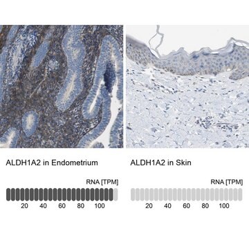
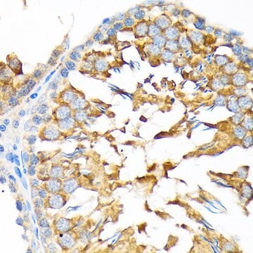
Anti-RalDH2 (ALDH1A2) Antibody, Cat. No. ABN420, is a highly specific rabbit polyclonal antibody that targets RalDH2 (ALDH1A2) and has been tested in Immunofluorescence, Immunohistochemistry (Paraffin), and Western Blotting.
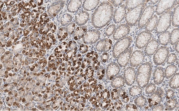
Immunohistochemistry Analysis: A 1:1,000 dilution from a representative lot detected RalDH2 in rat embryo tissue section.

Immunofluorescence Analysis: A 1:300 dilution from a representative lot immunostained the developing eye in paraffin-embedded embryonic E15 Sprague Dawley (SD) rat head sections by fluorescent immunohistochemistry (Courtesy of Anna Ashton, University of Aberdeen).

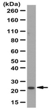
Western Blotting Analysis: A 1:3,000 dilution from a representative lot detected RalDH2 in 50 µg of new born (postnatal P0) Sprague Dawley (SD) rat whole eye tissue lysate, but not in adult SD rat brain lateral cortex and pons tissue lysates (Courtesy of Anna Ashton, University of Aberdeen).
Research Category
Neuroscience

Quality

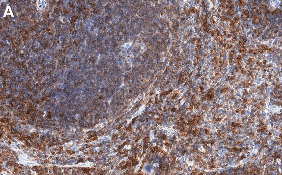
Evaluated by Immunohistochemistry in mouse retina tissue section.

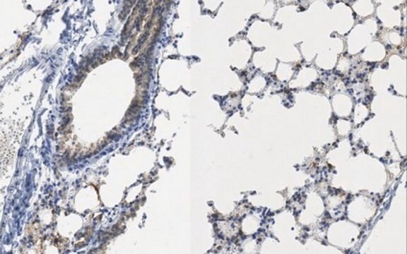
Immunohistochemistry Analysis: A 1:250 dilution of this antibody detected RalDH2 in mouse retina tissue section.

Target description

~57 kDa observed. 56.72/56.63/56.64 kDa (human/mouse/rat) calculated. Uncharacterized bands may be observed in some lysate(s).

Physical form

Affinity purified.
Purified rabbit polyclonal antibody in buffer containing 0.1 M Tris-Glycine (pH 7.4), 150 mM NaCl with 0.05% sodium azide.

Storage and Stability

Stable for 1 year at 2-8°C from date of receipt.

Other Notes

Concentration: Please refer to lot specific datasheet.

Disclaimer

Unless otherwise stated in our catalog or other company documentation accompanying the product(s), our products are intended for research use only and are not to be used for any other purpose, which includes but is not limited to, unauthorized commercial uses, in vitro diagnostic uses, ex vivo or in vivo therapeutic uses or any type of consumption or application to humans or animals.

Not finding the right product?  

Try our Product Selector Tool.

Storage Class

12 - Non Combustible Liquids

wgk_germany

WGK 1

flash_point_f

Not applicable

flash_point_c

Not applicable


Certificates of Analysis (COA)

Search for Certificates of Analysis (COA) by entering the products Lot/Batch Number. Lot and Batch Numbers can be found on a product’s label following the words ‘Lot’ or ‘Batch’.

Already Own This Product?

Find documentation for the products that you have recently purchased in the Document Library.

Visit the Document Library

Thabat Khatib et al.
Journal of Alzheimer's disease : JAD, 73(3), 935-954 (2019-12-31)
Retinoic acid has been previously proposed in the treatment of Alzheimer's disease (AD). Here, five transgenic mouse models expressing AD and frontotemporal dementia risk genes (i.e., PLB2APP, PLB2TAU, PLB1Double, PLB1Triple, and PLB4) were used to investigate if consistent alterations exist
Fabio Da Silva et al.
eLife, 10 (2021-10-09)
Retinoic acid (RA) is an essential signaling molecule for cardiac development and plays a protective role in the heart after myocardial infarction (MI). In both cases, the effect of RA signaling on cardiomyocytes, the principle cell type of the heart

Our team of scientists has experience in all areas of research including Life Science, Material Science, Chemical Synthesis, Chromatography, Analytical and many others.

Contact Technical Service