
C2724
Anti-Calbindin-D-28K (EG-20) antibody produced in rabbit
affinity isolated antibody, buffered aqueous solution
Synonym(s):
Anti-CALB, Anti-D-28K
Sign Into View Organizational & Contract Pricing
Select a Size
All Photos(3)
Select a Size
Change View
About This Item
Recommended Products


biological source
rabbit
Quality Level
conjugate
unconjugated
antibody form
affinity isolated antibody
antibody product type
primary antibodies
clone
polyclonal
form
buffered aqueous solution
mol wt
antigen 28 kDa
species reactivity
rat
enhanced validation
independent
Learn more about Antibody Enhanced Validation
General description
Calbindin-D-28K (CALB1) is a calcium binding protein in the brain. This 27kDa protein is located on human chromosome 8q21.
Calbindin-D-28k is an intracellular protein characterized by EF-hand type structural motifs that bind to Ca2+. This protein modulates synaptic plasticity and signal transduction . Anti-Calbindin-D-28K (EG-20) antibody recognizes rat calbindin-D-28K (28 kDa). Due to interspecies similarity, reactivity with human, mouse, bovine, chicken, porcine, and frog is expected, but has not been confirmed. Staining of calbindin-D-28K is specifically inhibited with the calbindin-D-28K immunizing peptide by immunoblotting.
Immunogen
The immunogen sequence is specific for calbindin-D-28K and is not found in other members of the EF-hand family such as calbindin-D-9K, calretinin, myosin light chain, parvalbumin, S-100a, S-100b, S100A2 (S100L) and S100A6 (calcyclin). This sequence is highly conserved across species, including human, mouse, bovine, chicken, porcine, and frog calbindin-D-28K sequences.
synthetic peptide corresponding to the C-terminal region of rat calbindin-D-28K (amino acids 225-244 with N-terminally added Lys-Gly) conjugated to KLH.
Application
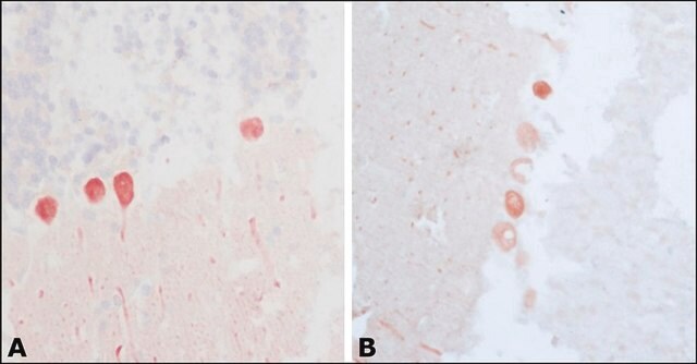
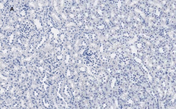
Anti-Calbindin-D-28K (EG-20) antibody is suitable for use in immunohistochemistry (1:2,000 using sections of rat cerebellum), immunoblot and western blot (1:2,000 using a whole extract of rat brain). Enzymatic predigestion of formalin-fixed, paraffin-embedded sections by proteolytic enzymes (e.g., 0.1% trypsin or protease, 10 min., at RT or 37 ° C) improves immunohistochemical staining with the antibody.
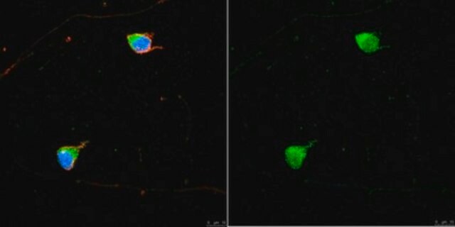
Anti-Calbindin-D-28K (EG-20) antibody has been used in immunohistochemical (IHC) peroxidase staining and cupric silver staining. It has also been used in immunofluorescence.
Anti-Calbindin-D-28K (EG-20) antibody has been used in immunohistochemical (IHC) peroxidase staining and cupric silver staining. It has also been used in immunofluorescence.
Physical form
Solution in 0.01 M phosphate buffered saline, pH 7.4, containing 1% bovine serum albumin and 15 mM sodium azide.
Analysis Note
Enzymatic predigestion of formalin-fixed, paraffin-embedded sections by proteolytic enzymes (e.g., 0.1% trypsin or protease, 10 min at RT or 37 °C) improves immunohistochemical staining.
Disclaimer
Unless otherwise stated in our catalog or other company documentation accompanying the product(s), our products are intended for research use only and are not to be used for any other purpose, which includes but is not limited to, unauthorized commercial uses, in vitro diagnostic uses, ex vivo or in vivo therapeutic uses or any type of consumption or application to humans or animals.
Not finding the right product?
Try our Product Selector Tool.
recommended
Product No.
Description
Pricing
Storage Class Code
12 - Non Combustible Liquids
WGK
nwg
Flash Point(F)
Not applicable
Flash Point(C)
Not applicable
Choose from one of the most recent versions:
Already Own This Product?
Find documentation for the products that you have recently purchased in the Document Library.
Customers Also Viewed

Postnatal phencyclidine administration selectively reduces adult cortical parvalbumin-containing interneurons
Wang C Z, et al.
Neuropsychopharmacology, 33(10), 2442-2442 (2008)
Long-term decrease in calbindin-D28K expression in the hippocampus of epileptic rats following pilocarpine-induced status epilepticus
Carter D S, et al.
Epilepsy Research, 79(2-3), 213-223 (2008)
Vascular endothelial growth factor increases during blood-brain barrier-enhanced permeability caused by Phoneutria nigriventer spider venom
Mendonca, M. C, et al.
BioMed Research International, 2014(6), 414-414 (2014)
GABA and synaptic inhibition of mouse cerebellum lacking glutamate decarboxylase 67
Obata K, et al.
Biochemical and Biophysical Research Communications, 370(3), 429-433 (2008)
Vaibhav P Pai et al.
Development (Cambridge, England), 139(2), 313-323 (2011-12-14)
Uncovering the molecular mechanisms of eye development is crucial for understanding the embryonic morphogenesis of complex structures, as well as for the establishment of novel biomedical approaches to address birth defects and injuries of the visual system. Here, we characterize
Our team of scientists has experience in all areas of research including Life Science, Material Science, Chemical Synthesis, Chromatography, Analytical and many others.
Contact Technical Service