Skip to Content
Merck
All Photos(1)

Documents

M5815

Sigma-Aldrich

Monoclonal Anti-Myogenin antibody produced in mouse

clone F12B, purified immunoglobulin, buffered aqueous solution

Sign Into View Organizational & Contract Pricing


About This Item

MDL number:
UNSPSC Code:
12352203
NACRES:
NA.41

biological source

mouse

Quality Level

conjugate

unconjugated

antibody form

purified immunoglobulin

antibody product type

primary antibodies

clone

F12B, monoclonal

form

buffered aqueous solution

mol wt

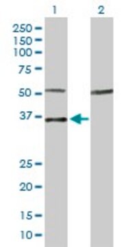
antigen 34 kDa

species reactivity

mouse, rat, human

technique(s)

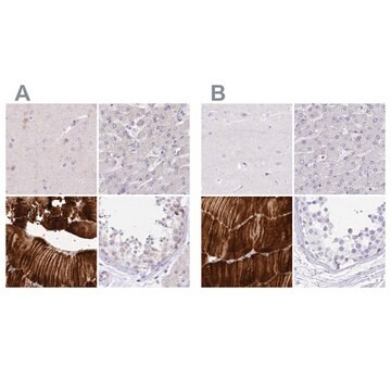
immunohistochemistry (formalin-fixed, paraffin-embedded sections): 1-2 μg/mL using rhabdomyosarcoma tissue sections
immunoprecipitation (IP): 2 μg using 1 mg protein lysate
indirect ELISA: suitable
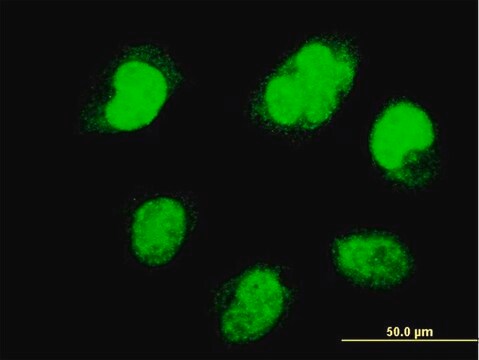
indirect immunofluorescence: suitable
western blot: 1 μg/mL

isotype

IgG1

UniProt accession no.

shipped in

wet ice

storage temp.

−20°C

target post-translational modification

unmodified

Gene Information

human ... MYOG(4656)
mouse ... Myog(17928)
rat ... Myog(29148)

Related Categories

General description

Myogenin belongs to the basic helix-loop-helix (bHLH) superfamily.
Myogenin is a transcription factor present in the nucleus. It possesses an amino-acid rich region and a helix-loop-helix structure. The gene encoding it is localized on human chromosome 1q31-41 and has three exons.

Immunogen

rat recombinant myogenin (amino acids 30-224).

Application

Monoclonal Anti-Myogenin antibody produced in mouse has been used in immunohistochemistry, immunofluorescence and western blotting.

Biochem/physiol Actions

Myogenin acts as a biomarker in rhabdomyosarcoma (RMS). It has a role in developmental processes, such as hematopoiesis, neurogenesis, myogenesis, tumor cell invasion and metastasis.
Myogenin acts as a regulator during myoblast and skeletal muscle differentiation. It is considered as a definite marker for skeletal muscle differentiation.

Physical form

Solution in phosphate buffered saline containing 0.08% sodium azide.

Disclaimer

Unless otherwise stated in our catalog or other company documentation accompanying the product(s), our products are intended for research use only and are not to be used for any other purpose, which includes but is not limited to, unauthorized commercial uses, in vitro diagnostic uses, ex vivo or in vivo therapeutic uses or any type of consumption or application to humans or animals.

Not finding the right product?  

Try our Product Selector Tool.

Storage Class Code

10 - Combustible liquids

WGK

nwg

Flash Point(F)

Not applicable

Flash Point(C)

Not applicable


Certificates of Analysis (COA)

Search for Certificates of Analysis (COA) by entering the products Lot/Batch Number. Lot and Batch Numbers can be found on a product’s label following the words ‘Lot’ or ‘Batch’.

Already Own This Product?

Find documentation for the products that you have recently purchased in the Document Library.

Visit the Document Library

Dysregulated Myogenesis in Rhabdomyosarcoma
Current Topics in Developmental Biology, 126(9), 285-297 (2018)
H Shen et al.
Brazilian journal of medical and biological research = Revista brasileira de pesquisas medicas e biologicas, 49(2), e5124-e5124 (2016-02-13)
This study evaluated the effect of muscle satellite cells (MSCs) overexpressing myogenin (MyoG) on denervated muscle atrophy. Rat MSCs were isolated and transfected with the MyoG-EGFP plasmid vector GV143. MyoG-transfected MSCs (MTMs) were transplanted into rat gastrocnemius muscles at 1
Neuraminidase-1 mediates skeletal muscle regeneration
de Carvalho Neves J, et al.
Biochimica et Biophysica Acta (BBA)-Molecular Basis of Disease, 1852(9), 1755-1764 (2015)
A Pertille et al.
Brazilian journal of biology = Revista brasleira de biologia, 77(1), 83-91 (2016-07-08)
The aim of this study was to analyze muscle regeneration after cryoinjury in the tibialis anterior muscle of young rats that were malnourished and then recovered. Forty Wistar rats were divided into a nourished group that received a normal protein
Hideki Ohnota et al.
Biochemical and biophysical research communications, 528(2), 398-403 (2020-01-14)
We have established a novel, simple, and highly reproducible method to generate skeletal muscle cells from mouse skin. Small pieces of skin from the back of mice were cultured in extracellular material-coated dishes in typical culture medium for about 3

Our team of scientists has experience in all areas of research including Life Science, Material Science, Chemical Synthesis, Chromatography, Analytical and many others.

Contact Technical Service