05-1143
Anti-phospho-Paxillin (Tyr31) Antibody, clone M102
clone M102, from mouse
Sign Into View Organizational & Contract Pricing
All Photos(1)
About This Item
UNSPSC Code:
12352203
eCl@ss:
32160702
NACRES:
NA.41
Recommended Products
biological source
mouse
Quality Level
antibody form
purified antibody
antibody product type
primary antibodies
clone
M102, monoclonal
species reactivity
human
technique(s)
western blot: suitable
isotype
IgG1
UniProt accession no.
shipped in
wet ice
target post-translational modification
phosphorylation (pTyr319)
Gene Information
human ... PXN(5829)
General description
Paxillin is involved in focal adhesion formation during cell adhesion and migration. Paxillin contains LD motifs, LIM domains, and an SH3- and SH2-binding domain that participate in a variety of protein-protein interactions with kinases, GTPase-activating proteins, and cytoskeletal proteins. Phosphorylation of paxillin occurs at both tyrosine and serine sites. Tyrosine phosphorylation of paxillin occurs in response to growth factors, neuropeptides, and integrins. The major sites of tyrosine phosphorylation include Tyr31 and Tyr118. Both of these sites may be involved in Crk binding to paxillin during integrin-mediated cell adhesion. These sites may provide docking motifs for recruitment of other signaling molecules to focal adhesions.
Specificity
The antibody detects Paxillin phosphorylated at Tyr31.
Immunogen
Clone M102 was generated from phospho-Paxillin (Tyr31) synthetic peptide (coupled to KLH) corresponding to amino acid residues around tyrosine 31 of human paxillin. This human sequence is highly conserved in rat and mouse paxillin.
Application
Detect phospho-Paxillin (Tyr31) using this Anti-phospho-Paxillin (Tyr31) Antibody, clone M102 validated for use in WB.
Research Category
Cell Structure
Cell Structure
Research Sub Category
Cytoskeletal Signaling
Cytoskeletal Signaling
Quality
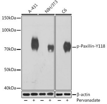
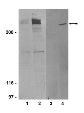
Evaluated by western Blot in pervanadate treated L6 cell lysates.
Western Blot Analysis: 1:500 dilution of this lot detected phospho-Paxillin on 10 µg of pervanadate treated L6 lysates.
Western Blot Analysis: 1:500 dilution of this lot detected phospho-Paxillin on 10 µg of pervanadate treated L6 lysates.
Target description
72 kDa
Linkage
Replaces: MAB1146
Physical form
Format: Purified
Protein A chromatography
Purified monoclonal IgG1 in phosphate-buffered saline, 50% glycerol, 1 mg/mL BSA, and 0.05% sodium azide.
Storage and Stability
Maintain at -20°C for one year from date of receipt. Do not aliquot.
Note: Variability in freezer temperatures below -20°C may cause glycerol containing solutions to become frozen during storage.
Note: Variability in freezer temperatures below -20°C may cause glycerol containing solutions to become frozen during storage.
Analysis Note
Control
Pervanadate treated L6 cell lysates
Pervanadate treated L6 cell lysates
Other Notes
Concentration: Please refer to the Certificate of Analysis for the lot-specific concentration.
Disclaimer
Unless otherwise stated in our catalog or other company documentation accompanying the product(s), our products are intended for research use only and are not to be used for any other purpose, which includes but is not limited to, unauthorized commercial uses, in vitro diagnostic uses, ex vivo or in vivo therapeutic uses or any type of consumption or application to humans or animals.
Not finding the right product?
Try our Product Selector Tool.
Storage Class Code
12 - Non Combustible Liquids
WGK
WGK 2
Flash Point(F)
Not applicable
Flash Point(C)
Not applicable
Certificates of Analysis (COA)
Search for Certificates of Analysis (COA) by entering the products Lot/Batch Number. Lot and Batch Numbers can be found on a product’s label following the words ‘Lot’ or ‘Batch’.
Already Own This Product?
Find documentation for the products that you have recently purchased in the Document Library.
Krista A Riggs et al.
Journal of cell science, 125(Pt 16), 3827-3839 (2012-05-11)
Integrins are the primary receptors of cells adhering to the extracellular matrix, and play key roles in various cellular processes including migration, proliferation and survival. The expression and distribution of integrins at the cell surface is controlled by endocytosis and
Our team of scientists has experience in all areas of research including Life Science, Material Science, Chemical Synthesis, Chromatography, Analytical and many others.
Contact Technical Service