Skip to Content
Merck
All Photos(2)

Key Documents

MABT126

Sigma-Aldrich

Anti-Obscurin Antibody, clone 5H10A

clone 5H10A, from mouse

Synonym(s):

EC:2.7.11.1, Obscurin-RhoGEF, Obscurin-myosin light chain kinase, Obscurin-MLCK

Sign Into View Organizational & Contract Pricing


About This Item

UNSPSC Code:
12352203
eCl@ss:
32160702

biological source

mouse

Quality Level

antibody form

purified immunoglobulin

antibody product type

primary antibodies

clone

5H10A, monoclonal

species reactivity

mouse, rat, human

technique(s)

immunofluorescence: suitable
immunohistochemistry: suitable
western blot: suitable

isotype

IgG1κ

NCBI accession no.

UniProt accession no.

shipped in

ambient

target post-translational modification

unmodified

Gene Information

human ... OBSCN(84033)

General description

Obscurin (UniProt: Q5VST9; also known as EC:2.7.11.1, Obscurin-RhoGEF, Obscurin-myosin light chain kinase, Obscurin-MLCK) is encoded by the OBSCN (also known as KIAA1556, KIAA1639) gene (Gene ID: 84033) in human. Obscurin is a member of the family of giant sarcomeric signaling proteins that includes titin and nebulin. It is expressed in cardiac and skeletal muscle, and plays a role in the organization of myofibrils during sarcomere assembly. Obscurin contains 68 Ig domains, 2 fibronectin domains, 1 calcium/calmodulin-binding domain, 1 RhoGEF domain with an associated PH domain, and 2 serine-threonine kinase domains. The dominant location of obscurin in mature myofibrils is shown to be at the peripheries of Z-disks and M-lines, where its position allows it to communicate with the surrounding myoplasm. Obscurin is reported to associates with the contractile apparatus through its binding to titin, sarcomeric myosin, and perhaps other proteins of the contractile apparatus. Obscurin is tightly associated with the mature contractile apparatus, hence it displays only limited solubility under non-denaturing conditions. However, it can be extracted from adult muscle fibers only in the presence of 2% SDS and 1% beta-mercaptoethanol. It is shown that primary cultures of neonatal skeletal myotubes contain large amounts of the 800 kDa obscurin isoform that can be solubilized with neutral detergents during early stages of sarcomere formation. (Ref.: Kontrogianni-Konstantopoulos, A., and Bloch, RJ. (2005). J Muscle Res Cell Motil. 26(6-8):419-26).

Specificity

Clone 5H10A detects obscurin in heart and skeletal muscle.

Immunogen

Epitope: N-terminus
GST-tagged full-length human recombinant Obscurin.

Application

Research Category
Cell Structure
This mouse monoclonal Anti-Obscurin, clone 5H10A Antibody, Cat. No. MABT126 is tested in Immunofluorescence Immunohistochemistry, and Western Blotting for the detection of obscurin.
Western Blotting Analysis: A representative lot detected Obscurin in Western Blotting applications (Bowman, A.L., et. al. (2007). FEBS Lett. 581(8):1549-54; Ackermann, M.A., et. al. (2014). PLoS One. 9(2):e88162).

Immunofluorescence Analysis: A representative lot detected Obscurin in Immunofluorescence applications (Bowman, A.L., et. al. (2007). FEBS Lett. 581(8):1549-54).

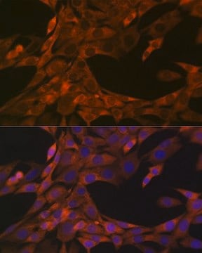
Immunofluorescence Analysis: A representative lot detected Obscurin in mouse heart left ventricle and mouse quadriceps muscle (Courtesy of Li-Yen R. Hu, Ph.D., and Aiketerini Kontrogianni-Konstantopoulos, Ph.D. , University of Maryland, School of Medicine, USA).

Immunohistochemistry Analysis: A representative lot detected Obscurin in Immunohistochemistry applications (Kontrogianni-Konstantopoulos, A., et. al. (2006). FASEB J. 20(12):2102-11; Ackermann, M.A., et. al. (2014). PLoS One. 9(2):e88162).

Western Blotting Analysis: A representative lot detected Obscurin in mouse heart left ventricle and mouse tibialis anterior muscle tissues (Courtesy of Li-Yen R. Hu, Ph.D., and Aiketerini Kontrogianni-Konstantopoulos, Ph.D. , University of Maryland, School of Medicine, USA).

Quality

Isotype testing: Identity Confirmation by Isotyping Test.

Isotyping Analysis: The identity of this monoclonal antibody is confirmed by isotyping test to be mouse IgG1.

Target description

~800 kDa observed; 868.48 kDa calculated. Uncharacterized bands may be observed in some lysate(s).

Physical form

Format: Purified
Protein G purified
Purified mouse monoclonal antibody IgG1 in buffer containing 0.1 M Tris-Glycine (pH 7.4), 150 mM NaCl with 0.05% sodium azide.

Storage and Stability

Stable for 1 year at 2-8°C from date of receipt.

Other Notes

Concentration: Please refer to lot specific datasheet.

Disclaimer

Unless otherwise stated in our catalog or other company documentation accompanying the product(s), our products are intended for research use only and are not to be used for any other purpose, which includes but is not limited to, unauthorized commercial uses, in vitro diagnostic uses, ex vivo or in vivo therapeutic uses or any type of consumption or application to humans or animals.

Not finding the right product?  

Try our Product Selector Tool.

Storage Class Code

12 - Non Combustible Liquids

WGK

WGK 1

Flash Point(F)

Not applicable

Flash Point(C)

Not applicable


Certificates of Analysis (COA)

Search for Certificates of Analysis (COA) by entering the products Lot/Batch Number. Lot and Batch Numbers can be found on a product’s label following the words ‘Lot’ or ‘Batch’.

Already Own This Product?

Find documentation for the products that you have recently purchased in the Document Library.

Visit the Document Library

Our team of scientists has experience in all areas of research including Life Science, Material Science, Chemical Synthesis, Chromatography, Analytical and many others.

Contact Technical Service