Skip to Content
Merck
All Photos(2)

Documents

16-222

Sigma-Aldrich

Anti-Phospho-Histone H3 (Ser10) Antibody, clone 3H10, FITC Conjugate

clone 3H10, Upstate®, from mouse

Synonym(s):

H3 histone, family 3A, H3S10P, Histone H3 (phospho S10), H3 histone, family 3A, H3 histone, family 3B, H3 histone, family 3B (H3.3B)

Sign Into View Organizational & Contract Pricing


About This Item

UNSPSC Code:
12352203
eCl@ss:
32160702
NACRES:
NA.41

biological source

mouse

Quality Level

conjugate

FITC conjugate

antibody form

purified antibody

antibody product type

primary antibodies

clone

3H10, monoclonal

species reactivity

human

manufacturer/tradename

Upstate®

technique(s)

immunocytochemistry: suitable
immunofluorescence: suitable
western blot: suitable

isotype

IgG

NCBI accession no.

UniProt accession no.

shipped in

wet ice

target post-translational modification

phosphorylation (pSer10)

Gene Information

human ... HIST1H3F(8968)

General description

Histone H3 is one of the five main histone proteins involved in the structure of chromatin in eukaryotic cells. Featuring a main globular domain and a long N-terminal tail, H3 is involved with the structure of the nucleosomes of the ′beads on a string′ structure. The N-terminal tail of histone H3 protrudes from the globular nucleosome core and can undergo several different types of epigenetic modifications that influence cellular processes. These modifications include the covalent attachment of methyl or acetyl groups to lysine and arginine amino acids and the phosphorylation of serine or threonine.

Specificity

Broad species cross-reactivity is expected.
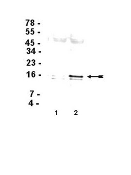
Recognizes histone H3 phosphorylated at Ser10, MW 17 kDa.

Immunogen

A proprietary immunogen based on a peptide sequence containing phospho-serine corresponding to residue 10 of human histone H3. Clone 3H10.
Epitope: Ser10

Application

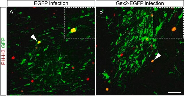
Anti-Phospho-Histone H3 (Ser10) Antibody, clone 3H10, FITC Conjugate is a Mouse Monoclonal for detection of Histone H3 phosphorylated at serine 10. This mAb, also known as H3S10p,is labeled with Fluorescein isothiocyanate (FITC), published in peer reviewed journals & validated in WB, ICC & IF.
Research Category
Epigenetics & Nuclear Function
Research Sub Category
Histones
Western Blot Analysis: 0.5-5 µg/mL of this lot detected phosphorylated histone H3 in acid extracted proteins from mitotic HeLa cells (Catalog # 17-306) treated with colcemid.

Quality

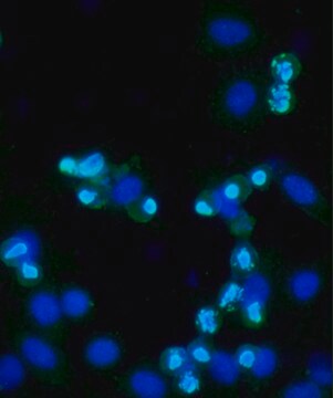
Immunocytochemistry: Mitotic HeLa cells showed positive chromosome staining with 4μg/mL of this antibody.

Target description

17 kDa

Physical form

Protein G Purified
Purified mouse monoclonal IgG in buffer containing PBS with 0.05% sodium azide, pH7.1.

Storage and Stability

Stable for 1 year at from date of receipt.

Analysis Note

Control
Mitotic HeLa cells (IF).

Legal Information

UPSTATE is a registered trademark of Merck KGaA, Darmstadt, Germany

Disclaimer

Unless otherwise stated in our catalog or other company documentation accompanying the product(s), our products are intended for research use only and are not to be used for any other purpose, which includes but is not limited to, unauthorized commercial uses, in vitro diagnostic uses, ex vivo or in vivo therapeutic uses or any type of consumption or application to humans or animals.

Not finding the right product?  

Try our Product Selector Tool.

Storage Class Code

10 - Combustible liquids

WGK

WGK 2


Certificates of Analysis (COA)

Search for Certificates of Analysis (COA) by entering the products Lot/Batch Number. Lot and Batch Numbers can be found on a product’s label following the words ‘Lot’ or ‘Batch’.

Already Own This Product?

Find documentation for the products that you have recently purchased in the Document Library.

Visit the Document Library

Tumor-suppressing function of caspase-2 requires catalytic site Cys-320 and site Ser-139 in mice.
Keqin Ren,Jing Lu,Aleksey Porollo,Chunying Du
The Journal of Biological Chemistry null
Antje M Wengner et al.
Molecular cancer therapeutics, 15(4), 583-592 (2016-02-03)
Monopolar spindle 1 (Mps1) has been shown to function as the key kinase that activates the spindle assembly checkpoint (SAC) to secure proper distribution of chromosomes to daughter cells. Here, we report the structure and functional characterization of two novel
Agata Zieba et al.
Molecular & cellular proteomics : MCP, 11(7), M111-M111 (2012-03-24)
Fundamental open questions in signal transduction remain concerning the sequence and distribution of molecular signaling events among individual cells. In this work, we have characterized the intercellular variability of transforming growth factor β-induced Smad interactions, providing essential information about TGF-β

Our team of scientists has experience in all areas of research including Life Science, Material Science, Chemical Synthesis, Chromatography, Analytical and many others.

Contact Technical Service