추천 제품
Quality Level
IVD
for in vitro diagnostic use
응용 분야
clinical testing
diagnostic assay manufacturing
hematology
histology
저장 온도
15-25°C
일반 설명
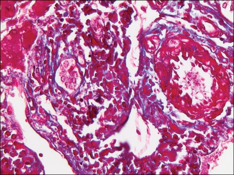
The Masson-Goldner staining kit - for the visualization of connective tissue with trichromic staining, is used for human-medical cell diagnosis and serves the purpose of the histological investigation of sample material of human origin. Using a combination of three different staining solutions, muscle fibers, collagenous fibers, fibrin and erythrocytes can be selectively visualized.
The original methods were primarily used to differentiate collagenous and muscle fibers. The stains used have different molecular sizes and enable the individual tissues to be stained differentially.
The Masson-Goldner staining technique can be carried out using formalin fixed material. Subsequent to staining the nucleus with Weigert′s iron hematoxylin, components such as muscle, cytoplasm and erythrocytes are stained with azophloxin and orange G solution. Connective tissue is then counter stained using light green SF solution.
The package is sufficient for 400 - 500 applications. The product is registered as IVD and CE product and can be used in diagnostics and laboratory accreditation. For more details, please see instructions for use (IFU). The IFU can be downloaded from this webpage.
분석 메모
Suitability for microscopy (Tissue section): passes test
Nuclei: dark brown to black
Cytoplasm: brick red
musculature (muscles): bright red
Connective tissue: green
acid mucosubstances: green
Erythrocytes: bright orange
Nuclei: dark brown to black
Cytoplasm: brick red
musculature (muscles): bright red
Connective tissue: green
acid mucosubstances: green
Erythrocytes: bright orange
신호어
Danger
유해 및 위험 성명서
Hazard Classifications
Eye Dam. 1 - Skin Irrit. 2
Storage Class Code
8A - Combustible, corrosive hazardous materials
WGK
WGK 2
시험 성적서(COA)
제품의 로트/배치 번호를 입력하여 시험 성적서(COA)을 검색하십시오. 로트 및 배치 번호는 제품 라벨에 있는 ‘로트’ 또는 ‘배치’라는 용어 뒤에서 찾을 수 있습니다.
Aurora Garre et al.
Clinical, cosmetic and investigational dermatology, 11, 253-263 (2018-06-09)
With age, decreasing dermal levels of proteoglycans, collagen, and elastin lead to the appearance of aged skin. Oxidation, largely driven by environmental factors, plays a central role. The aim of this study was to assess the antiaging efficacy of a
B Shen et al.
Nan fang yi ke da xue xue bao = Journal of Southern Medical University, 41(7), 1107-1113 (2021-07-27)
To investigate the effect of ginsenoside Rh2 (G-Rh2) on renal fibrosis and cell apoptosis in rats with diabetic nephropathy (DN) and explore its possible mechanism. Thirty male SD rats were randomized equally into control group, DN group and ginsenoside Rh2
Álvaro Santana-Garrido et al.
Journal of physiology and biochemistry (2022-08-10)
Arterial hypertension (AH) leads to oxidative and inflammatory imbalance that contribute to fibrosis development in many target organs. Here, we aimed to highlight the harmful effects of severe AH in the cornea. Our experimental model was established by administration of
자사의 과학자팀은 생명 과학, 재료 과학, 화학 합성, 크로마토그래피, 분석 및 기타 많은 영역을 포함한 모든 과학 분야에 경험이 있습니다..
고객지원팀으로 연락바랍니다.


