おすすめの製品
由来生物
mouse
品質水準
抗体製品の状態
purified immunoglobulin
抗体製品タイプ
primary antibodies
クローン
5F10, monoclonal
化学種の反応性
human
テクニック
flow cytometry: suitable
immunocytochemistry: suitable
inhibition assay: suitable
western blot: suitable
アイソタイプ
IgG2bλ
NCBIアクセッション番号
UniProtアクセッション番号
ターゲットの翻訳後修飾
unmodified
遺伝子情報
human ... F2RL3(9002)
詳細
Proteinase-activated receptor 4 (UniProt Q96RI0; also known as Coagulation factor II receptor-like 3, PAR-4, Thrombin receptor-like 3) is encoded by the F2RL3 (also known as PAR4) gene (Gene ID 9002) in human. Protease-activated receptors (PARs) constitute a unique family of seven-transmembrane, G-protein-coupled receptors (GPCRs) activated by proteolytic cleavage of their N-terminal propeptide sequence. Once cleaved off, the N-terminal propeptide fragment functions as a ligand and activates the receptor by binding the second extracellular loop. The four PAR family members (PAR-1 to PAR-4) are widely expressed and activated by multiple proteases, and utilize different types of G-proteins (Gi, Gq, and G12/13) for signal transdution depending on the activating protease and cellular context. PAR-4 is expressed on platelets and exhibits a low-affinity for thrombin. However, PAR-4 is able to form hetero-oligomers with both PAR-1 and the ADP receptor P2Y12 to mediate thrombin- and ADP-initiated signaling. PAR-4 cleavage is significantly enhanced through hetero-oligomerization with PAR-1, and PAR-4 interaction with P2Y12 is directly linked to arrestin-2 recruitment and AKT signaling. PAR-4 is a 7-transmembrane (a.a. 83-103, 109-129, 152-172, 192-213, 248-268, 284-304, 320-343) GPCR activated by thrombin cleavage between R47 and G48, having 3 extracellular loops and 3 intracellular loops between the extracellular N-terminal end (a.a. 48-82) the cytoplasmic C-terminal tail (a.a. 344-385).
特異性
Clone 5F10 recognizes a propeptide epitope near (N-terminal to) the thrombin-cleavage site and protects cell surface PAR4 against thrombin cleavage (Mumaw, M.M., et al. (2015). Thromb. Res. 135(6):1165-1171). Clone 5F10 recognizes prepro- and pro-, but not proteolytically activated, forms of human PAR4.
免疫原
Epitope: Near (N-terminal to) the thrombin cleavage site.
MBP-conjugated recombinant human PAR4 N-terminal fragment including the propeptide sequence.
アプリケーション
Research Category
細胞シグナル伝達
細胞シグナル伝達
Research Sub Category
GPCR、cAMP/cGMP及びカルシウムシグナル伝達
GPCR、cAMP/cGMP及びカルシウムシグナル伝達
Anti-PAR4 Antibody, clone 5F10 is an antibody against PAR4 for use in Western Blotting, Immunocytochemistry, Flow Cytometry, Inhibition.
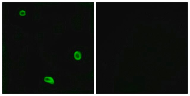
Immunocytochemistry Analysis: 20 µg/mL from a representative lot detected endogenous PAR4 by fluorescent immunocytochemistry staining of freshly isolated human platelets (Courtesy of Dr. Marvin Nieman, Case Western Reserve University, Cleveland, OH, USA).
Immunocytochemistry Analysis: A representative lot detected the expression of exogenously transfected human PAR4 by fluorescent immunocytochemistry staining of 4% formaldehyde-fixed HEK293 Flp-In cells following tetracycline treatment (Mumaw, M.M., et al. (2015). Thromb. Res. 135(6):1165-1171).
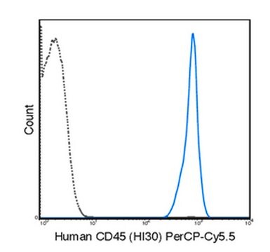
Flow Cytometry Analysis: A representative lot detected tetracycline-induced expression of exogenously transfected human PAR4 on the surface of HEK293 Flp-In cells. Thrombin treatment diminished cell surface PAR4 immunoreactivity (Mumaw, M.M., et al. (2015). Thromb. Res. 135(6):1165-1171).
Inhibition Analysis: A representative lot, when added prior to thrombin, protected cell surface PAR4 against thrombin cleavage (Mumaw, M.M., et al. (2015). Thromb. Res. 135(6):1165-1171).
Western Blotting Analysis: A representative lot detected MBP fusion proteins containing human PAR4 fragment a.a. 18-78 or 41-66, but not 48-72. MBP-PAR4 fusion degradation by thrombin treatment ablolished target band detection by clone 5F10 (Mumaw, M.M., et al. (2015). Thromb. Res. 135(6):1165-1171).
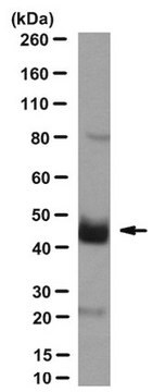
Western Blotting Analysis: A representative lot detected tetracycline-induced expression of exogenously introduced human PAR4 in a HEK293 Flp-In cell line, as well as endogenous PAR4 in isolated human platelets (hPLTs). Thrombin activation of hPLTs diminished PAR4 target band detection (Mumaw, M.M., et al. (2015). Thromb. Res. 135(6):1165-1171).
品質
Evaluated by Western Blotting in human platelet lysate.
Western Blotting Analysis: A 1:250 dilution of this antibody detected PAR4 in 50 µg of human platelet lysate.
Western Blotting Analysis: A 1:250 dilution of this antibody detected PAR4 in 50 µg of human platelet lysate.
ターゲットの説明
~45 kDa observed. 41.13/39.16 kDa (prepro-/pro-PAR4) calculated. The broad banding pattern and larger apparent band size is consistent with the detection of glycosylated PAR4. Uncharacterized band(s) may appear in some lysates.
物理的形状
Protein G purified.
Format: Purified
Purified mouse monoclonal IgG2bλ antibody in PBS without preservatives.
保管および安定性
Stable for 1 year at -20°C from date of receipt.
その他情報
Concentration: Please refer to lot specific datasheet.
免責事項
Unless otherwise stated in our catalog or other company documentation accompanying the product(s), our products are intended for research use only and are not to be used for any other purpose, which includes but is not limited to, unauthorized commercial uses, in vitro diagnostic uses, ex vivo or in vivo therapeutic uses or any type of consumption or application to humans or animals.
適切な製品が見つかりませんか。
製品選択ツール.をお試しください
保管分類コード
12 - Non Combustible Liquids
WGK
WGK 2
引火点(°F)
Not applicable
引火点(℃)
Not applicable
適用法令
試験研究用途を考慮した関連法令を主に挙げております。化学物質以外については、一部の情報のみ提供しています。 製品を安全かつ合法的に使用することは、使用者の義務です。最新情報により修正される場合があります。WEBの反映には時間を要することがあるため、適宜SDSをご参照ください。
Jan Code
MABS1298:
試験成績書(COA)
製品のロット番号・バッチ番号を入力して、試験成績書(COA) を検索できます。ロット番号・バッチ番号は、製品ラベルに「Lot」または「Batch」に続いて記載されています。
ライフサイエンス、有機合成、材料科学、クロマトグラフィー、分析など、あらゆる分野の研究に経験のあるメンバーがおります。.
製品に関するお問い合わせはこちら(テクニカルサービス)


