Passa al contenuto
Merck
Tutte le immagini(8)

Documenti fondamentali

P0372

Sigma-Aldrich

Anti-Podocin antibody produced in rabbit

affinity isolated antibody, buffered aqueous solution

Sinonimo/i:

Podocin Antibody, Podocin Antibody - Anti-Podocin antibody produced in rabbit

Autenticatiper visualizzare i prezzi riservati alla tua organizzazione & contrattuali

Scegli un formato


Scegli un formato

Cambia visualizzazione

About This Item

Numero MDL:
Codice UNSPSC:
12352203
NACRES:
NA.41

Origine biologica

rabbit

Livello qualitativo

Coniugato

unconjugated

Forma dell’anticorpo

affinity isolated antibody

Tipo di anticorpo

primary antibodies

Clone

polyclonal

Stato

buffered aqueous solution

PM

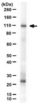
antigen ~42 kDa (doublet)

Reattività contro le specie

human, rat, mouse

tecniche

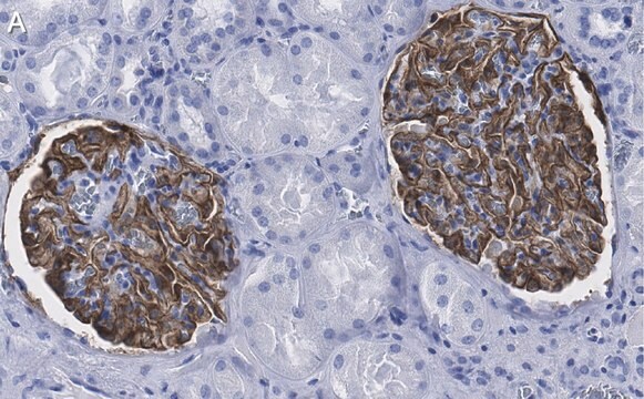
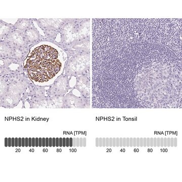
indirect immunofluorescence: 10-20 μg/mL using acetone-fixed human or rat kidney frozen sections
western blot (chemiluminescent): 0.5-1 μg/mL using whole extract of rat glomeruli

Descrizione generale

Podocin is a podocyte structural protein, that belongs to the band-7-stomatin family of lipid raft-associated proteins. This family contains more than 1800 conserved proteins which share ∼150 amino acid domains showing similarity to mitochondrial protein prohibitin (PHB).
Podocin is a hairpin-like integral membrane protein with intracellular N- and C- termini. Podocin is located at the insertion site of the slit membrane, an intercellular junction found in mammalian kidney.

Specificità

Anti-Podocin recognizes human, mouse, and rat podocin.

Immunogeno

Synthetic peptide corresponding to amino acid residues of human podocin with an N-terminal added cysteine, conjugated to KLH. The corresponding sequence is identical in mouse and differs by one amino acid in rat.

Applicazioni

Anti-Podocin antibody produced in rabbit has been used for Western Blotting and immunohistochemistry.

Azioni biochim/fisiol

Podocin and nephrin are essential for maintaining the slit diaphragm. Studies show that mutations in this gene account for around 15% cases of nephrotic syndrome cases. Podocin interacts with cholesterol and promotes the formation of protein-lipid supercomplexes, which control ion channel complexes. The podocin gene is known to be associated with autosomal recessive SRNS (nephrotic syndrome type 2). Podocin actively participates in the ultrafiltration process occuring in kidney.

Stato fisico

Solution in 0.01 M phosphate buffered saline, pH 7.4, containing 15 mM sodium azide.

Stoccaggio e stabilità

For continuous use, store at 2-8 °C for up to one month.
For extended storage, freeze in working aliquots. Repeated freezing and thawing is not recommended. Storage in frost-free freezers is also not recommended. If slight turbidity occurs upon prolonged storage, clarify the solution by centrifugation before use. Working dilutions should be discarded if not used within 12 hours.

Esclusione di responsabilità

Unless otherwise stated in our catalog or other company documentation accompanying the product(s), our products are intended for research use only and are not to be used for any other purpose, which includes but is not limited to, unauthorized commercial uses, in vitro diagnostic uses, ex vivo or in vivo therapeutic uses or any type of consumption or application to humans or animals.

Non trovi il prodotto giusto?  

Prova il nostro Motore di ricerca dei prodotti.

Codice della classe di stoccaggio

12 - Non Combustible Liquids

Classe di pericolosità dell'acqua (WGK)

nwg

Punto d’infiammabilità (°F)

Not applicable

Punto d’infiammabilità (°C)

Not applicable


Scegli una delle versioni più recenti:

Certificati d'analisi (COA)

Lot/Batch Number

Non trovi la versione di tuo interesse?

Se hai bisogno di una versione specifica, puoi cercare il certificato tramite il numero di lotto.

Possiedi già questo prodotto?

I documenti relativi ai prodotti acquistati recentemente sono disponibili nell’Archivio dei documenti.

Visita l’Archivio dei documenti

I clienti hanno visto anche

Slide 1 of 3

1 of 3

The SPFH domain-containing proteins: more than lipid raft markers.
Browman D T, et al.
Trends in Cell Biology, 17(8), 394-402 (2007)
Macrophages directly mediate diabetic renal injury.
You H et al
American Journal of Physiology: Renal Physiology, 305(12), F1719-F1727 (2013)
Bart Smeets et al.
Journal of the American Society of Nephrology : JASN, 20(12), 2604-2615 (2009-11-18)
Cellular lesions form in Bowman's space in both crescentic glomerulonephritis and collapsing glomerulopathy. The pathomechanism and origin of the proliferating cells in these lesions are unknown. In this study, we examined proliferating cells by lineage tracing of either podocytes or
Induction of podocyte VEGF164 overexpression at different stages of development causes congenital nephrosis or steroid-resistant nephrotic syndrome.
Veron D et al
The American Journal of Pathology, 177(5), 2225-2233 (2010)
Batoul Wehbi et al.
Journal of the American Society of Nephrology : JASN, 30(7), 1238-1249 (2019-06-23)
IgA nephropathy (IgAN) often follows infections and features IgA mesangial deposition. Polymeric IgA deposits in the mesangium seem to have varied pathogenic potential, but understanding their pathogenicity remains a challenge. Most mesangial IgA1 in human IgAN has a hypogalactosylated hinge

Il team dei nostri ricercatori vanta grande esperienza in tutte le aree della ricerca quali Life Science, scienza dei materiali, sintesi chimica, cromatografia, discipline analitiche, ecc..

Contatta l'Assistenza Tecnica.