PKH and CellVue® Claret Fluorescent Cell Linker Dyes
PKH and CellVue® Fluorescent Cell Linker Kits provide fluorescent labeling of live cells over an extended period of time, with no apparent toxic effects. Fluorescent linker kits are effective for a variety of cell types and exhibit no significant leaking, or transfer from cell to cell. They provide stable, clear, intense, accurate and reproducible fluorescent labeling of cells. Patented cell linker technology incorporates aliphatic reporter molecules into the cell membrane lipid bilayer by selective partitioning.
Four fluorescent linkers are available. PKH2 and PKH67 are green fluorochromes with excitation (490 nm) and emission (504 nm) similar to fluorescein, while PKH26 is a red fluorochrome, has excitation (551 nm) and emission (567 nm) characteristics compatible with rhodamine or phycoerythrin detection systems. PKH26 may also be excited by the 488 nm emission of an argon-ion laser. CellVue® Claret is a far-red fluorochrome which is independent of pH in physiologic ranges, and has excitation (655 nm) and emission (675 nm) characteristics compatible with a red diode laser. The linkers are physiologically stable and show little to no toxic side-effects on cell systems. Labeled cells retain both biological and proliferative activity, and are ideal for cell tracking and cell-cell interaction studies.
Due to the non-specific labeling mechanism of the cell linkers, a wide variety of cell types have been labeled successfully. The linkers have been applied to both animal and plant cells as well as other membrane containing particles. The pattern of staining is dependent upon the cell type being labeled and the membrane of the cells. Although most applications center around general labeling (GL) methods involving membrane incorporation of the probes, the linkers may also be used for selective phagocytic cell labeling (PCL). Appearance of labeled cells may vary from bright "immunofluorescence" labeling to a punctate or patchy appearance. Since the labeling is not a saturation reaction, but rather a function of both dye and cell concentration, it is essential that the amount of dye available for incorporation be limited. Overlabeling of the cells will result in loss of membrane integrity and cell recovery.
Features:
- Fluorescent labeling of live cells
- Stable for up to 100 days on live cells in vivo
- Intense and easy-to-use
- Non-cytotoxic - no effect on biological or proliferative activity
- Versatile - labels various eukaryotic cells and cell lines, bacteria, and parasites
- Uniform and reproducible
- No significant dye leakage or transfer from cell to cell
- Different dyes permit multi-color analysis
- Compatible with other fluorescent labels
- Technology that works, with hundreds of literature references to prove it
View the JoVE video: Optimized Staining and Proliferation Modeling Methods for Cell Division Monitoring using Cell Tracking Dyes
Use of PKH and CellVue® dyes to monitor cell trafficking and function: Bibliography
Comparison of CFSE and PKH26 with CellVue® Claret, A New 675 nm-emitting Proliferation Dye
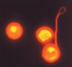
Figure 2. Mouse Hematopoietic Stem Cells Stained with PKH26
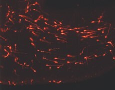
Figure 3. Young Migrating Neurons Labeled with PKH26
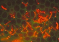
Figure 4. Plasmodium Stained with PKH26

Figure 5.Confocal Assay for Phagocytosis
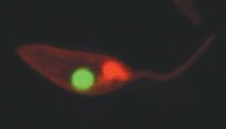
Figure 6. Leishmania donovani Labeled with PKH26
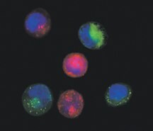
Figure 7.Coculture of Chemosensitive and Chemoresistant Labeled T24 Cells

Figure 8.PKH67 in L929 Cells
To continue reading please sign in or create an account.
Don't Have An Account?