Novel and Rapid Method to Assess Proliferative Properties of Mammalian Cells by Monitoring Cell Size Distributions
Abstract
Much research has recently focused on elucidating the mechanisms by which eukaryotic cells regulate their size with respect to growth rate and cell cycle stage. These mechanisms are known to differ between yeast and higher eukaryotes, and may also differ between cell types. However, because of the challenges of isolating individual factors affecting cell size, and because of the challenges of measuring cell size distributions quickly and precisely, the signaling pathways governing mammalian cell size are still unknown (Tzur, 2010). In this study, we have employed impedance-based volume/size determination and flow cytometric analysis to investigate the relationship between cell cycle, cell proliferation rate, and cell size.
By arresting cultured cells at various stages of the cell cycle, we demonstrated that multiple types of cells undergo changes in cell size with respect to cell cycle. Further, our data indicated that the cell size distribution of exponentially growing cells (both adherent and suspension) is reproducible, independent of passage number (Halter, 2008). The size distribution represents the relative cell number in each phase of the cell cycle. We showed, using flow cytometry, that the constant shape of the size distribution between passages represented constant percentages of the different phases of the cell cycle.
Finally, we monitored the size distributions of several cell lines (both adherent and suspension) over a 7-day period. We documented a shift of the size distribution from larger, exponentially growing cells toward smaller, G0/G1 phase cells. We could confirm that this shift of the cell size represented a shift in the ratio of the different phases towards an increasing percentage of cells in the G0/G1 phase and a decrease in division rate. Again, the shape of this cell size distribution, when majority of cells are in the G0/G1 phase, was reproducible between passages. Our results indicate that monitoring the shape of the cell size distribution is a fast and useful method for assessing the proliferative properties of mammalian cells.
Introduction
We have employed the Scepter system, a handheld, automated cell counter that instantly reports cell size/volume distributions as well as a Guava® benchtop microcapillary flow cytometer, to investigate the relationship between cell cycle, cell proliferation rate, and cell size/volume. Our results show that monitoring the shape of the cell size/volume distribution is a useful method for a rapid QC assessment of your cell culture (apoptosis, cell cycle disruption and proliferation). Further in depth detailed analysis can be acquired afterwards using Guava.
Materials & Methods
Cells and media:
M4A4 (ATCC Catalog No. CRL2914) and NIH3T3 (ATCC Catalog No. CRL1658) were maintained in DMEM (Catalog No. SLM-021-B)+ 10% FBS (Catalog No. ES-009-B) + 1% Glutamax™ supplement (Life Technologies, Catalog No. 35050) + 1% Penicillin Streptomycin (GIBCO Catalog No. 15140). Jurkat (ATCC Catalog No. TIB 152) were cultured in RPMI 1640 (Catalog No. SLM 140-B) + 10% FBS.
Instruments and reagents:
Scepter™ Cell Counter, Scepter Software 1.2 (Catalog No. PHC00000) and Scepter sensors 60 µM (Catalog No. PHCC60050)
Guava EasyCyte™ 8HT Flow Cytometer
Guava Nexin® Reagent (Catalog No. 4500-0450)
Cell Cycle Analysis Assay (Catalog No. 4500-0220)
Colchicine (Catalog No. C9754)
Camptothecin (Catalog No. 208925)
Sample preparation and analysis:
For experiments, cells were seeded in 6-well plate, each time three replicates were used per data point. For each data point, cells were harvested and pelleted. Pellets were resuspended in PBS and cell suspensions were counted using Scepter device. Data was downloaded and analyzed using Scepter Software 1.2 and histograms were generated using averages of three replicates. For further analysis with Guava (Nexin, Cell Cycle) reagents and assays same samples were processed via assay’s instructions.
Scepter™ System: rapid assessment of cell size changes
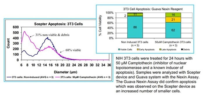
Tracking apoptosis
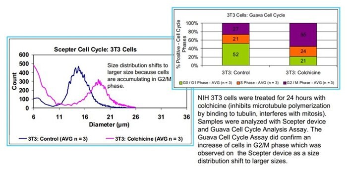
Tracking cell cycle disruption
Tracking proliferative properties
Exponential phase: Reproducible cell size distributions of exponentially growing cells independent of passage number.

Cell counts and size distributions (average of n=3) from respectively M4A4 and Jurkat cells acquired with the Scepter device at different time points and analyzed using Scepter Software 1.2.
End of exponential phase: decreasing peak size value predicts the end of exponential growth.
U266: suspension cell line, human myeloma cells
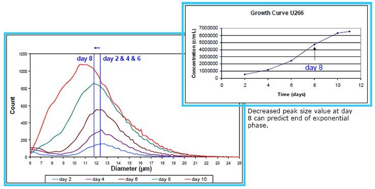
Cell counts and size distributions were taken every day (n=3) over an 11 day period usting the Scepter device. Data were analyzed using Scepter Software 1.2. Peak size values decreased at day 8.
M4A4: adherent cell line, human breast carcinoma cells
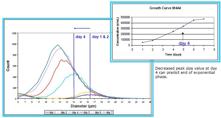
Cell counts and size distributions were taken every day (n=3) over a 7 day period with the Scepter device. Data were analyzed using Scepter Software 1.2. Peak size values decreased at day 4.
M4A4 cells: Guava Cell Cycle
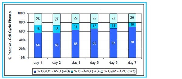
The same M4A4 samples were analyzed with Guava Cell Cycle Assay. Day 1 & 2 with similar peak size showed similar % of the different cell phase. The decrease in peak size value correspond to an increasing % of cells in G0/G1 and a slight decrease in % of cells in S and G2/M phases.
Scepter Device: rapid QC assessment of cell culture
Histograms of M4A4 culture, displayed with Scepter Software 1.2.

Conclusion
- The Scepter device can be used as a cell culture monitoring device for a qualitative assessment of apoptosis and cell cycle disruption. More detailed analysis can be performed using Guava flow cytometer.
- Monitoring cell size distributions is indicative of the proliferative properties of your cells.
- In general, the Scepter device can be used for a rapid QC assessment of your cell culture.
References
To continue reading please sign in or create an account.
Don't Have An Account?