Skip to Content
Merck
All Photos(3)

Documents

HPA001171

Sigma-Aldrich

Anti-PLA2G6 antibody produced in rabbit

enhanced validation

Prestige Antibodies® Powered by Atlas Antibodies, affinity isolated antibody, buffered aqueous glycerol solution

Synonym(s):

Anti-NBIA2, Anti-PARK14, Anti-PNPLA9, Anti-iPLA2, Anti-iPLA2beta, PLA2G6 Antibody - Anti-PLA2G6 antibody produced in rabbit, Pla2G6 Antibody

Sign Into View Organizational & Contract Pricing


About This Item

MDL number:
UNSPSC Code:
12352203
Human Protein Atlas Number:
NACRES:
NA.41

biological source

rabbit

conjugate

unconjugated

antibody form

affinity isolated antibody

antibody product type

primary antibodies

clone

polyclonal

product line

Prestige Antibodies® Powered by Atlas Antibodies

form

buffered aqueous glycerol solution

species reactivity

human

enhanced validation

orthogonal RNAseq
recombinant expression
Learn more about Antibody Enhanced Validation

technique(s)

immunoblotting: 0.04-0.4 μg/mL
immunofluorescence: 0.25-2 μg/mL

immunogen sequence

QVLHTEVLQHLTDLIRNHPSWSVAHLAVELGIRECFHHSRIISCANCAENEEGCTPLHLACRKGDGEILVELVQYCHTQMDVTDYKGETVFHYAVQGDNSQVLQLLGRNAVAGLNQVNNQGLTPLHLACQLGKQEMVRVLLLCNARCNIMG

UniProt accession no.

shipped in

wet ice

storage temp.

−20°C

target post-translational modification

unmodified

Gene Information

human ... PLA2G6(8398)

General description

PLA2G6 (phospholipase A2, group VI) localizes in the mitochondria.

Immunogen

85 kDa calcium-independent phospholipase A2 recombinant protein epitope signature tag (PrEST)

Application

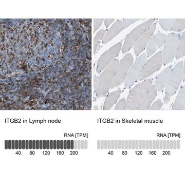
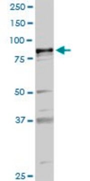
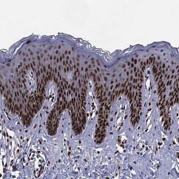
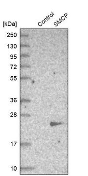
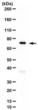
Anti-PLA2G6 antibody produced in rabbit, a Prestige Antibody, is developed and validated by the Human Protein Atlas (HPA) project . Each antibody is tested by immunohistochemistry against hundreds of normal and disease tissues. These images can be viewed on the Human Protein Atlas (HPA) site by clicking on the Image Gallery link. The antibodies are also tested using immunofluorescence and western blotting. To view these protocols and other useful information about Prestige Antibodies and the HPA, visit sigma.com/prestige.

Biochem/physiol Actions

PLA2G6 (phospholipase A2, group VI) gene encodes a phospholipase that catalyzes the release of fatty acids from phospholipids. The protein may function in phospholipid remodeling, nitric oxide-induced or vasopressin-induced arachidonic acid release. It is also involved in leukotriene and prostaglandin synthesis. It may have a role in fas mediated apoptosis and in regulating transmembrane ion flux in glucose-stimulated B-cells. It is also involved in cardiolipin (CL) deacylation. Mutations in PLA2G6 are associated with infantile neuroaxonal dystrophy, neurodegeneration with brain iron accumulation type II and autosomal recessive early-onset dystonia-parkinsonism. Loss of PLA2G6 gene activity causes lipid peroxidation, mitochondrial dysfunction and thereby mitochondrial membrane abnormalities. It is also involved in type 1 diabetes, by participating in endoplasmic reticulum stress-induced β-cell apoptosis.

Features and Benefits

Prestige Antibodies® are highly characterized and extensively validated antibodies with the added benefit of all available characterization data for each target being accessible via the Human Protein Atlas portal linked just below the product name at the top of this page. The uniqueness and low cross-reactivity of the Prestige Antibodies® to other proteins are due to a thorough selection of antigen regions, affinity purification, and stringent selection. Prestige antigen controls are available for every corresponding Prestige Antibody and can be found in the linkage section.

Every Prestige Antibody is tested in the following ways:
  • IHC tissue array of 44 normal human tissues and 20 of the most common cancer type tissues.
  • Protein array of 364 human recombinant protein fragments.

Linkage

Corresponding Antigen APREST73385

Physical form

Solution in phosphate-buffered saline, pH 7.2, containing 40% glycerol and 0.02% sodium azide

Legal Information

Prestige Antibodies is a registered trademark of Merck KGaA, Darmstadt, Germany

Disclaimer

Unless otherwise stated in our catalog or other company documentation accompanying the product(s), our products are intended for research use only and are not to be used for any other purpose, which includes but is not limited to, unauthorized commercial uses, in vitro diagnostic uses, ex vivo or in vivo therapeutic uses or any type of consumption or application to humans or animals.

Not finding the right product?  

Try our Product Selector Tool.

Storage Class Code

10 - Combustible liquids

WGK

WGK 1

Personal Protective Equipment

dust mask type N95 (US), Eyeshields, Gloves

Certificates of Analysis (COA)

Search for Certificates of Analysis (COA) by entering the products Lot/Batch Number. Lot and Batch Numbers can be found on a product’s label following the words ‘Lot’ or ‘Batch’.

Already Own This Product?

Find documentation for the products that you have recently purchased in the Document Library.

Visit the Document Library

François Gonzalvez et al.
Biochimica et biophysica acta, 1832(8), 1194-1206 (2013-03-26)
Cardiolipin is a mitochondrion-specific phospholipid that stabilizes the assembly of respiratory chain complexes, favoring full-yield operation. It also mediates key steps in apoptosis. In Barth syndrome, an X chromosome-linked cardiomyopathy caused by tafazzin mutations, cardiolipins display acyl chain modifications and
Yuan-Hao Hsu et al.
Journal of the American Chemical Society, 135(4), 1330-1337 (2012-12-22)
The mechanism of inhibition of group VIA Ca(2+)-independent phospholipase A(2) (iPLA(2)) by fluoroketone (FK) ligands is examined by a combination of deuterium exchange mass spectrometry (DXMS) and molecular dynamics (MD). Models for iPLA(2) were built by homology with the known
Xiaoyong Lei et al.
Endocrinology, 155(9), 3352-3364 (2014-07-09)
Type 1 diabetes (T1D) results from autoimmune destruction of islet β-cells, but the underlying mechanisms that contribute to this process are incompletely understood, especially the role of lipid signals generated by β-cells. Proinflammatory cytokines induce ER stress in β-cells and
Xiaoyong Lei et al.
American journal of physiology. Endocrinology and metabolism, 303(11), E1386-E1395 (2012-10-18)
Death of β-cells due to apoptosis is an important contributor to β-cell dysfunction in both type 1 and type 2 diabetes mellitus. Previously, we described participation of the Group VIA Ca(2+)-independent phospholipase A(2) (iPLA(2)β) in apoptosis of insulinoma cells due
A Paliege et al.
American journal of physiology. Renal physiology, 302(7), F865-F874 (2012-01-06)
Na(+)-K(+)-2Cl(-) cotransporter (NKCC2)-mediated NaCl reabsorption in the thick ascending limb (TAL) is stimulated by AVP via V2 receptor/PKA/cAMP signaling. This process is antagonized by locally produced eicosanoids such as 20-HETE or prostaglandin E(2), which are synthesized in a phospholipase A(2)-dependent

Our team of scientists has experience in all areas of research including Life Science, Material Science, Chemical Synthesis, Chromatography, Analytical and many others.

Contact Technical Service