MABT329M
Anti-Cytokeratin 8 Antibody, clone TROMA-1
clone TROMA-1, from rat
Synonym(s):
Keratin type II cytoskeletal 8, Cytokeratin endo A, Cytokeratin-8, CK-8, Keratin-8, K8, Type-II keratin Kb8
About This Item
Recommended Products
biological source
rat
Quality Level
antibody form
purified immunoglobulin
antibody product type
primary antibodies
clone
TROMA-1, monoclonal
species reactivity
human, mouse
packaging
antibody small pack of 25 μg
technique(s)
immunocytochemistry: suitable
immunofluorescence: suitable
immunohistochemistry: suitable (paraffin)
immunoprecipitation (IP): suitable
radioimmunoassay: suitable
western blot: suitable
isotype
IgG2aκ
NCBI accession no.
UniProt accession no.
target post-translational modification
unmodified
Gene Information
human ... KRT8(3856)
mouse ... Krt8(16691)
General description
Specificity
Immunogen
Application
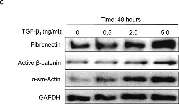
Western Blotting Analysis: A representative lot detected Cytokeratin 8 in Western Blotting applications (Helenius, T.O., et. al. (2016). Cells. 5(3); O′Driscoll, C.M., et. al. (2013). Biochim Biophys Acta. 1833(3):460-7; Tao, G.Z., et. al. (2003). J Cell Sci. 116(Pt 22):4629-38; Maurer, J., et. al. (2008) PLoS One. 3(10):e3451).
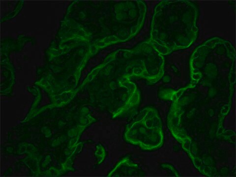
Immunocytochemistry Analysis: A 1:250 dilution from a representative lot detected Cytokeratin 8 in HepG2 cells.
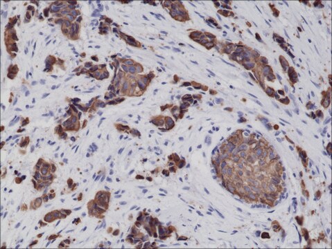
Immunohistochemistry Analysis: A 1:50 dilution from a representative lot detected Cytokeratin 8 in mouse lung tissue.
Immunoprecipitation Analysis: A representative lot detected Cytokeratin 8 in Immunoprecipitation applications (Brulet, P., et. al. (1982). Proc Natl Acad Sci USA. 79(7):2328-32).
Radioimmunoassay Analysis: A representative lot detected Cytokeratin 8 in Radioimmunoassy applications (Brulet, P., et. al. (1982). Proc Natl Acad Sci USA. 79(7):2328-32).
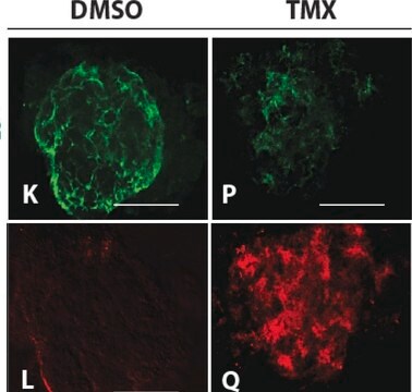
Immunofluorescence Analysis: A representative lot detected Cytokeratin 8 in Immunofluorescence applications (Helenius, T.O., et. al. (2016). Cells. 5(3); Tao, G.Z., et. al. (2003). J Cell Sci. 116(Pt 22):4629-38; Lin, Y.M., et. al. (2008). J Cell Sci. 121(Pt 14):2382-93; Maurer, J., et. al. (2008) PLoS One. 3(10):e3451; Nuotio-Antar, A.M., et. al. (2006). J Steroid Biochem Mol Biol. 99(2-3):93-9; Tamai, Y., et. al. (2000). J Cell Biol. 151(3):563-72).
Immunohistochemistry Analysis: A representative lot detected Cytokeratin 8 in Immunohistochemistry applications (Szeder, V., et. al. (2003). Dev Biol. 253(2):258-63; Tao, G.Z., et. al. (2003). J Cell Sci. 116(Pt 22):4629-38; Nuotio-Antar, A.M., et. al. (2006). J Steroid Biochem Mol Biol. 99(2-3):93-9; Tamai, Y., et. al. (2000). J Cell Biol. 151(3):563-72).
Quality
Western Blotting Analysis: 0.5 ug/mL of this antibody detected Cytokeratin 8 in 10 µg of HeLa cell lysate.
Target description
Physical form
Other Notes
Not finding the right product?
Try our Product Selector Tool.
Storage Class Code
12 - Non Combustible Liquids
WGK
WGK 1
Certificates of Analysis (COA)
Search for Certificates of Analysis (COA) by entering the products Lot/Batch Number. Lot and Batch Numbers can be found on a product’s label following the words ‘Lot’ or ‘Batch’.
Already Own This Product?
Find documentation for the products that you have recently purchased in the Document Library.
Our team of scientists has experience in all areas of research including Life Science, Material Science, Chemical Synthesis, Chromatography, Analytical and many others.
Contact Technical Service